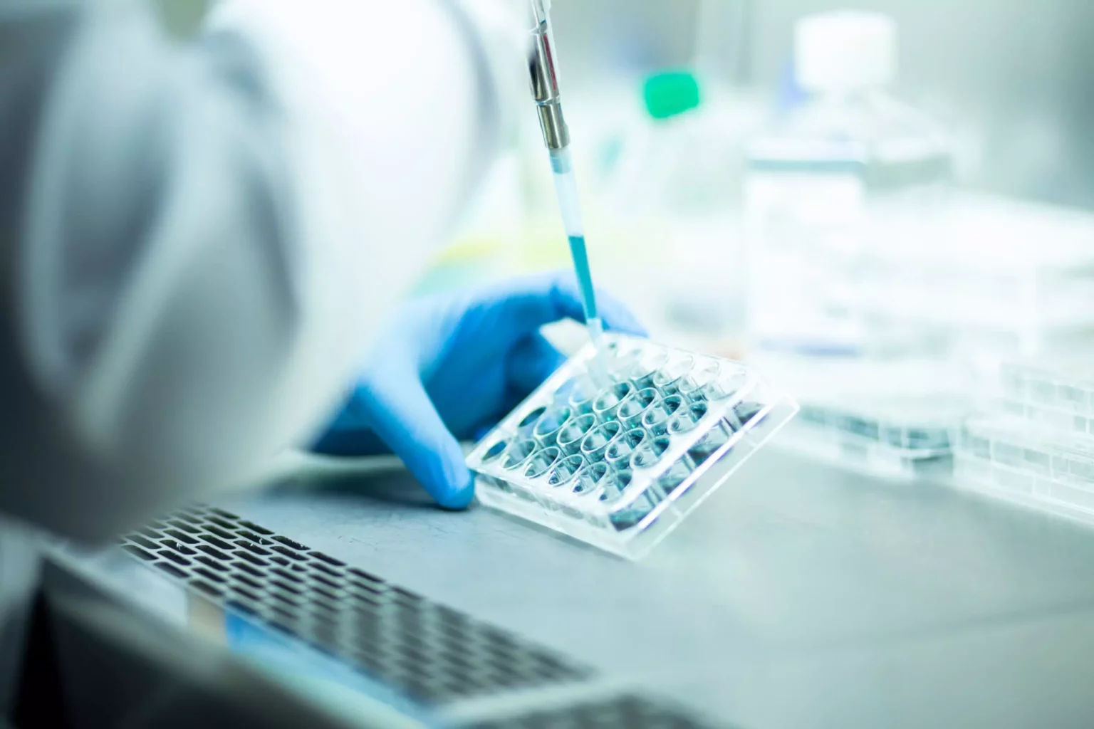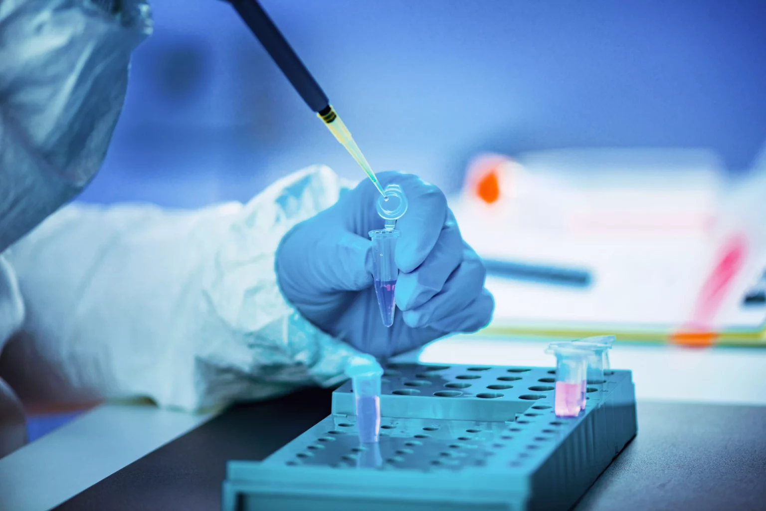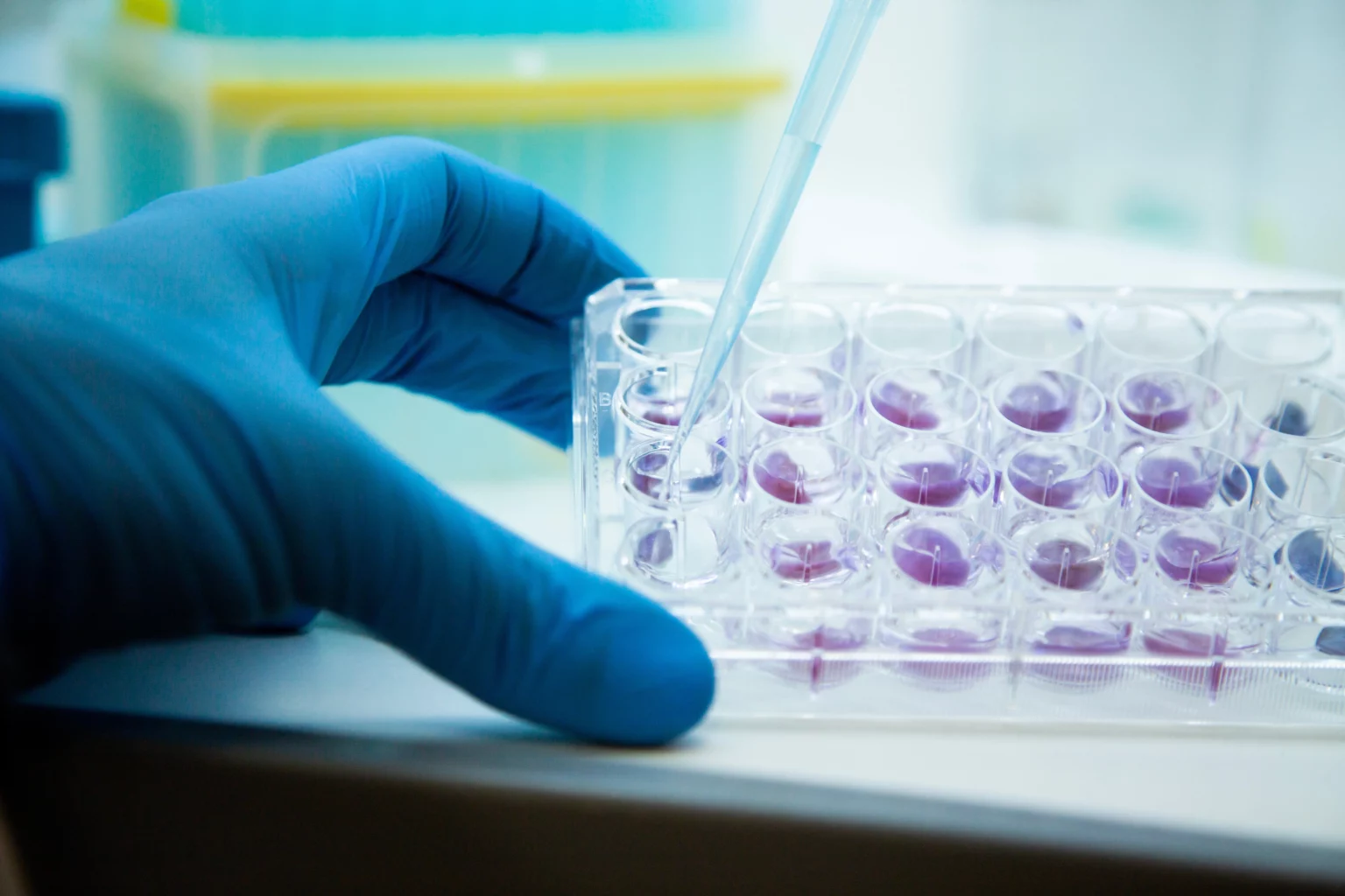
The first of our fields of application is cytology. The ScreenCell Cyto device is dedicated to cytology techniques: cytomorphology, immunocytology and 2D or 3D FISH.
The morphology of the fixed cells retained on the membrane is well preserved. The filters can be observed under a microscope immediately after staining with a nucleo-cytoplasmic dye.
The same filters can then be destained and immunolabeled, allowing correlation with the morphologically identified cells.
The cells are analyzed according to classic malignancy criteria in order to distinguish normal leukocytes from atypical cells consistent with the characteristics of circulating tumor cells (CTCs).
In addition, ScreenCell technology makes it possible to identify CTCs clusters as well as large atypical macrophages called CAMLs (Cancer-Associated Macrophage-Like) and platelets agglomerated with CTCs clusters in microemboli.
Iterative counts of CTCs and CTC clusters are useful for monitoring patients after treatment. This would make it possible to detect a relapse as early as possible even if there is not yet a detectable tumor.
Iterative immunostaining is useful for searching for new therapeutic directions that may be different from those identified through primary tumor biopsy.
For molecular characterization of CTCs, blood must be filtered using the ScreenCell MB kit. At the end of the filtration process, the live CTCs are retained on the isolation support, ready to be lysed directly in the capsule. Thus, the MB capsule opens access to the genome and transcriptome of CTCs and allows a wide range of applications.
The ScreenCell MB kit is compatible with various DNA/RNA extraction kits. Do not hesitate to ask us for more details on the protocols.
In addition, ScreenCell technology also makes it possible to study CTCs DNA and circulating DNA from the same blood sample in parallel.


ScreenCell technology helps preserve the viability as well as the proliferative and invasive potential of CTCs. The cells of interest are isolated alive then placed in culture, thus allowing the expansion and ex-vivo characterization of CTCs subpopulations. The optimal technical and micro-environmental conditions for the isolation and expansion of CTCs have been defined for ScreenCell devices. This gives us opportunities to understand the process of metastasis and drug resistance.
Alternatively, cells can be detached from the membrane after isolation and obtained in suspension to subsequently enable the analysis of individual cells as well as other applications such as 3D culture.
Automated page speed optimizations for fast site performance