Optimizing Detection of Circulating Tumor Cells in Breast Cancer: Unveiling New Markers for Clinical Applications
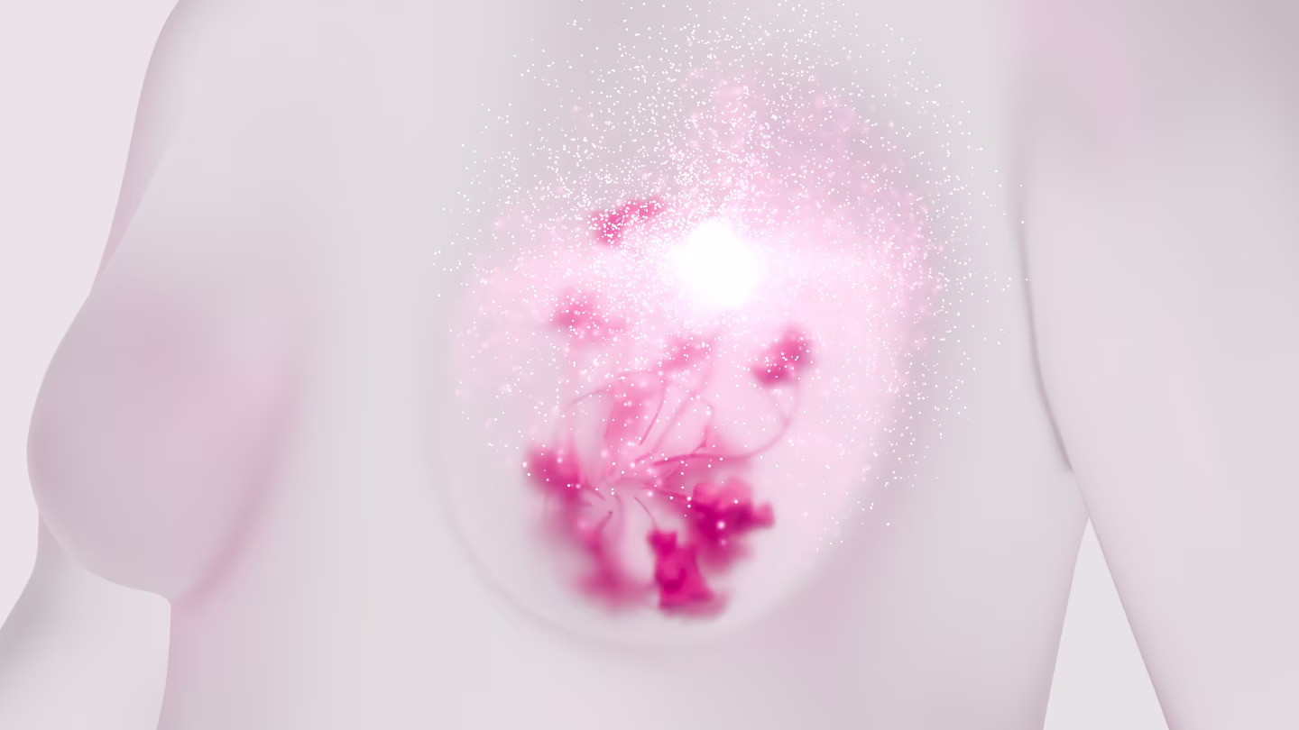
Breast cancer (BC) is a heterogeneous disease with high metastasis potential, especially in the bones, liver, and lungs. Circulating tumor cells (CTCs), which emerge from active tumors, represent an early step toward metastasis and are associated with poor prognosis. CTCs of carcinoma origin are believed to express EpCAM and cytokeratins (CKs), common epithelial markers that are frequently used to identify them. However, in practice, the most aggressive CTCs lose the expression of those markers, leading to the partial loss of important information. Thus, finding some novel markers that identify CTCs regardless of their heterogeneity is crucial. A specific bioinformatics workflow integrating primary tumor and diverse BC cell lines transcriptomic expression analysis was developed and compared with single CTC transcriptomic analyses. We have identified a set of genes that are overexpressed in primary BC cells and are commonly upregulated among BC cell lines. Fifty of them were also found to be expressed in BC CTCs by single-cell transcriptomic analysis. Further in silico sorting narrowed this list to 12 genes. Using ScreenCell technology to isolate cancer cells spiked into normal blood, we tested the protein expression of all corresponding genes in vitro using the double immunocytochemistry method and validated MARCKSL1, SLC9A3R1, and RHOD as the most expressed markers. We then isolated the CTCs of 40 LN-invaded BC patients and 18 healthy donors using ScreenCell technology and showed that the combination of these three markers resulted in significantly better recognition of CTCs compared to EpCAM and CK conventional markers. Employing these novel markers, we found a clear distinction between blood samples from patients and healthy donors. In conclusion, through a specific bioinformatics workflow, in addition to in vitro and further clinical validations, we found three novel markers to precisely identify CTCs. These markers, when used together, enable a significantly more efficient identification of CTCs compared to conventional epithelial markers.
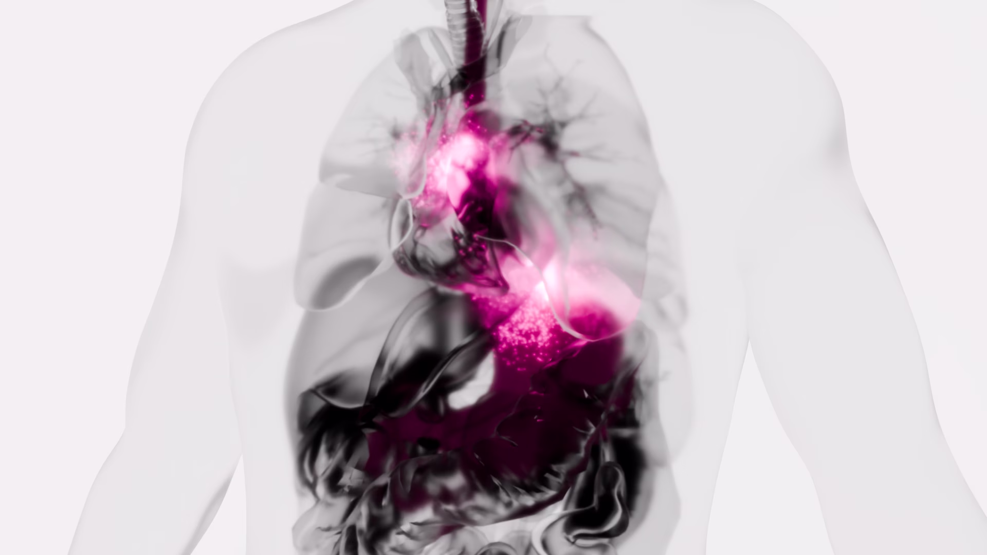
Circulating Tumor Cells from Surgical Manipulation Predict Recurrence and Poor Prognosis in Non-Small Cell Lung Cancer
Background/Objectives: In our previous multicenter prospective controlled study (UMIN000018602), we investigated the impact of surgical manipulation on circulating tumor cells (CTCs) in patients with non-small cell lung cancer (NSCLC). CTCs were detected after surgery in four patients (4/29, 13.8%), although CTCs were not present before surgery. These four patients had tumor cells leaked into their bloodstream by surgeons’ manipulation. We aimed to clarify long-term outcomes according to the presence of CTCs.
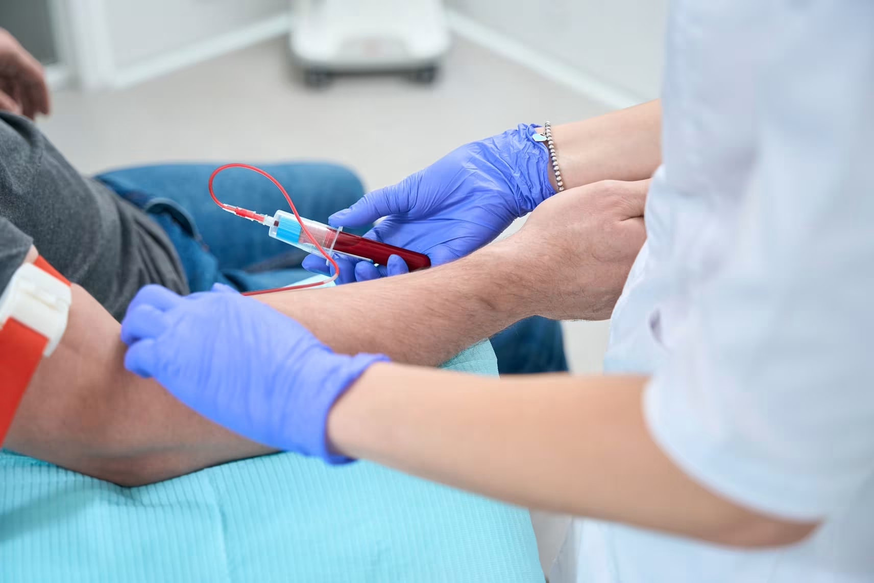
Methods: Patients with cT1b-2N0M0 NSCLC scheduled for lobectomy were enrolled, based on the selection criteria of a consolidation-to-ground-glass opacity ratio (over 50%). Peripheral blood samples (≥3 mL) were collected before surgery (for pre-CTCs), during surgery, and immediately after pulmonary vein dissection (for post-CTCs). CTCs were isolated from these samples using ScreenCell®’s size-selective method.
Results: From July 2015 to January 2016, 29 patients were enrolled, yielding paired pre- and post-CTC samples for all patients. Thirteen patients were pre-CTC positive, and post-CTCs were detected in 17 patients. Survival analysis revealed a statistically significant difference in recurrence-free survival between patients with and without post-CTCs (p = 0.043), while pre-CTCs status had no significant impact on recurrence (p = 0.226). Patients with post-CTCs had a significantly higher recurrence rate than those without (p = 0.043). Half of patients with post-CTCs but without pre-CTCs had recurrence within 5 years after surgery.
Conclusions: Post-CTCs emerged as a significant predictor of recurrence following lobectomy; however, it could be possible for thoracic surgeons to prevent recurrence by improving surgical techniques for NSCLC patients with post-CTCs but without pre-CTCs.
Optimum diagnostic pathway and pathologic confirmation rate of early stage lung cancer: Results from the VIOLET randomised controlled trial
Background: Pathologic confirmation of lung cancer influences treatment selection for suspected early-stage lung cancer. High pre-treatment tissue confirmation rates are recommended. We sought to define management and outcomes of patients undergoing surgery for primary lung cancer in a UK multi-centre clinical trial.
Methods: VIOLET compared minimally invasive video-assisted thoracic surgery versus open surgery for known or suspected lung cancer. Diagnostic patient pathways were identified and methods of tissue confirmation were documented. The outcome of inappropriate lobectomy for benign disease or inappropriate wedge resection for primary lung cancer was compared with respect to the pathologic diagnosis.
Findings: From July 2015 to February 2019, 502 patients were randomised and underwent surgery; 262 (52%) had a pre-operative pathologic confirmed diagnosis of primary lung cancer, 205 did not have a pre-operative biopsy and 35 had a non-diagnostic pre-operative biopsy. Of the 240 participants without pre-operative pathologic confirmation of primary lung cancer, intraoperative biopsy and frozen section analysis was undertaken in 144 (60%). The remaining 96 underwent direct surgical resection without tissue confirmation (19% of the entire cohort). Confirmation of histologic diagnosis before surgery was less costly than diagnosis in the operating theatre. The inappropriate surgery rate was 3.6% (18/502 participants, 7 lobectomy for benign disease, 11 wedge resection for lung cancer).
Interpretation: Low levels of inappropriate resection can be achieved at pre-operative tissue confirmation rates of 50% through a combination of intra-operative confirmatory biopsy and correct risk estimation of lung cancer. Practice needs to be monitored to ensure acceptable levels are consistently achieved.
Fast and efficient isolation of murine circulating tumor cells using screencell technology for pre-clinical analyzes
Circulating tumor cells (CTCs) represent a rare and heterogeneous population of cancer cells that are detached from the tumor site and entered blood or lymphatic circulation. Once disseminated in distant tissues, CTCs could remain dormant or create a tumor mass causing serious danger for patients. Many technologies exist to isolate CTCs from patients’ blood samples, mostly based on microfluidic systems or by sorting them according to their surface antigens, notably EpCAM, and/or cytokeratins for carcinoma. ScreenCell has developed an easy-to-use, antigen-independent, rapid, cost-effective, and efficient technology that isolates CTCs according to their bigger size compared to the blood cells. This study provides the technical information necessary to isolate and characterize CTCs from mouse blood. By using blood samples from transgenic mice with breast cancer or from WT mice in which we spiked cancer cells, we showed that ScreenCell technology is compatible with standard EDTA blood collection tubes. Furthermore, the ScreenCell Cyto kit could treat up to 500 µl and the ScreenCell MB kit up to 200 µl of mouse blood. As the ScreenCell MB kit captures unaltered live CTCs, we have shown that their DNA could be efficiently extracted, and the isolated cells could be grown in culture. In conclusion, ScreenCell provides a rapid, easy, antigen-independent, cost-effective, and efficient technology to isolate and characterize CTCs from the blood samples of cancer patients and murine models. Thanks to this technology CTCs could be captured fixed or alive. Murine cancer models are extensively used in pre-clinical studies. Therefore, this study demonstrates the crucial technical points necessary while manipulating mouse blood samples using ScreenCell technology.
An assessment of diagnostic performance of a filter-based antibody-independent peripheral blood circulating tumour cell capture paired with cytomorphologic criteria for the diagnosis of cancer
Objectives
Circulating tumour cells (CTCs) are reported to be predictive for prognosis and response to treatment in advanced lung cancer. However, the clinical utility of the CTCs detection remains unknown for early stage lung cancer as the number of CTCs is reported as low, providing challenges in identification. We have evaluated diagnostic performance of filtration-based technology using cytomorphologic criteria in patients undergoing surgery for lung cancer.
Material and methods
We processed blood from 76 patients undergoing surgery for known or suspected lung cancer using ScreenCell® Cyto filter devices. Captured cells were stained using haematoxylin and eosin and independently assessed by two pathologists for the presence of atypical cells suspicious for cancer. Diagnostic performance was evaluated against pathologist reported diagnoses of cancer from surgically obtained specimens.
Results
Cancer was diagnosed in 57 patients (77.0%), including 32 with primary lung cancer (56.1%). The proportion of patients with early stage primary lung cancer in which CTCs were identified was 18 and 21 (56.3% and 65.6%, respectively) as reported by two pathologists. The agreement between the pathologists was 77.0% corresponding to a kappa-statistic of 53.7% indicating moderate agreement. No significant differences were found for the percentage of CTCs for primary and metastatic cancer as well as for cancer stages. On sensitivity weighted analysis, a sensitivity and specificity were 71.9% (95% CI 60.5–83.0) and 52.9% (95% CI 31.1–77.0), respectively. On specificity weighted analysis, a sensitivity and specificity were 50.9% (95% CI 39.3–64.4) and 82.4% (60.4–96.2), respectively.
Conclusion
The performance of the tested filter-based antibody-independent technology to capture CTCs using standard cytomorphologic criteria provides the potential of a diagnostic blood test for lung cancer.
Evaluating circulating tumour cell enrichment techniques to establish an appropriate method for clinical application in glioblastomas

Brain tumours reduce life expectancy for an average of 20 years per patient, the highest of any cancer. A third of brain tumour patients visit their GP at least five times before diagnosis and many of those are diagnosed late through emergency departments. A possible solution to this challenge is to utilise a “”liquid biopsy”” blood test designed for circulating tumour cells (CTCs). Such a test could be applied at a primary healthcare centre, contributing to informed decision making for diagnostic imaging referrals. Furthermore, it could also be applied at secondary health care centres for the ongoing monitoring of disease recurrence. There is increased interest in CTC enrichment methods as a potential approach for faster diagnosis and monitoring of disease progression. The aim of this review to compare four CTC enrichment methods – OncoQuick®, Screen Cell®, pluriBead® and Cell Search® – with the objective of identifying a suitable method for application in the clinical setting for the isolation of CTCs from glioblastomas.
Cancer-associated Macrophage-like Cells in Patients with Non-metastatic Adenocarcinoma of the Esophagus – Cytomorphological Heterogeneity
Introduction: Esophageal adenocarcinoma (EAC) often recurs systemically despite therapy with a curative aim. New diagnostic and therapeutic approaches are urgently needed. A promising field is liquid biopsy, meaning the investigation of tumor-associated cells in the peripheral blood, for example cancer-associated macrophage-like cells (CAML). The aim of this multicentric study was to investigate the presence and cytomorphological appearance of CAML in patients with non-metastatic and operable esophageal cancer.
Methods: Blood samples from 252 patients with locally advanced EAC were obtained before starting curative treatment including surgery, and then processed using ScreenCell® filtration devices. Cytological analysis was performed via May-Grünwald-Giemsa staining. CAML were defined by their morphological characteristics. We also performed immunofluorescence staining with the mesenchymal marker vimentin on a subset of our study cohort.
Results: We detected cytomorphologically heterogeneous CAML in 31.8% (n=80) patients. Their presence and cell count did not correlate significantly with pretherapeutic cTNM. Even in patients with small tumors and no lymph-node infiltration, cell counts were high. CAML showed heterogenous staining patterns for vimentin.
Conclusion: This is one of the first studies demonstrating the presence and phenotype of CAML in a uniquely broad cohort of EAC patients. As they are believed to be representatives of the inflammatory tumor microenvironment shed into the bloodstream, their presence in non-metastatic EAC is a promising finding.
Cytomorphologic visualization of circulating tumor cells in urinary bladder cancer patients using ScreenCell™ technology: Potential as a simple cytology test

Circulating tumor cells (CTC) are a recent technique which is a potentially important prognostic factor in many solid tumors. There are many techniques of detecting CTCs, but they usually implement costly techniques like EpCAM targeted detection, fluorescence-based diagnosis, or magnetic bead based positive or negative selection. The diagnostic utility of simple cytomorphological diagnosis after routine staining of CTCs have been rarely studied. We aimed to detect CTCs in 24 patients clinically suspected to have Urinary Bladder Cancer using a simple but efficient patented filtration technology (ScreenCell™), followed by optical microscopic visualization after routine May-Grunwald-Giemsa (MGG) staining. The detected CTCs were then tested for association with the histologic type, lamina propria invasion, deep muscle invasion and the T-stage. Out of the 24 patients tested, one was found to have papilloma, nine had low grade urothelial carcinoma, 13 had high grade urothelial carcinoma and one had poorly differentiated adenocarcinoma. Of these, two LGUC, eight HGUC and one adenocarcinoma had detectable CTC. Presence of CTCs had a statistically significant association with Lamina propria invasion (P = .006) and T-stage (P = .02), and a trend toward significance for differentiating LGUC from HGUC (P = .10). These results suggest that cytomorphological detection of CTC is likely to be clinically useful in diagnosis and prognostication of urinary blader cancers. These findings need to be confirmed on studies with larger sample sizes.
Ex vivo expansion of circulating tumour cells (CTCs)
Circulating tumour cells (CTCs) are a critical intermediate step in the process of cancer metastasis. The reliability of CTC isolation/purification has limited both the potential to report on metastatic progression and the development of CTCs as targets for therapeutic intervention. Here we report a new methodology, which optimises the culture conditions for CTCs using primary cancer cells as a model system. We exploited the known biology that CTCs thrive in hypoxic conditions, with their survival and proliferation being reliant on the activation of hypoxia-inducible factor 1 alpha (HIF-1α). We isolated epithelial-like and quasi-mesenchymal CTC phenotypes from the blood of a cancer patient and successfully cultured these cells for more than 8 weeks. The presence of CTC clusters was required to establish and maintain long-term cultures. This novel methodology for the long-term culture of CTCs will aid in the development of downstream applications, including CTC theranostics.
A Fast and Furious Liquid Biopsy Assay to Monitor Targeted Therapy Resistance.
Liquid biopsies represent a valid alternative to conventional tissue biopsies, offering a real time molecular picture of tumors in a minimally invasive manner. Of the various circulating biomarkers available for liquid biopsy, circulating tumor cells (CTC) and circulating tumor DNA (ctDNA) are the most intensively studied to date. However, CTC and ctDNA represent different tumor components, therefore, complementary information from both sources might be beneficial. This protocol focuses on the description of a sample processing workflow that allowed for concurrent isolation of CTC and ctDNA from the same source sample. This single tube approach enables simultaneous analysis of multiple biomarkers to better monitor cancer drug resistance.