
Circulating Tumor Cells in Diagnosing Lung Cancer: Clinical and Morphologic Analysis

Background: The purpose of this study was to evaluate the value of circulating non-hematologic cells to differentiate benign from malignant lung lesions and their comparison with clinico-histologic features of corresponding primary lesions.
Methods: Circulating cells were isolated by size method from peripheral blood of 77 patients with malignant (n = 60) and benign (n = 17) lung lesions. They were morphologically classified as cells with malignant feature; cells with uncertain malignant feature; and cells with benign feature; then statistically correlated with clinico-cytopathologic characteristics of corresponding lung lesion.
Results: Malignant circulating cells were detected in 54 of 60 (90%) malignant patients, and in 1 of 17 (5%) benign patients; benign circulating cells in 1 of 60 (1%) malignant patients and in 15 of 17 (88%) benign patients; and circulating cells with uncertain malignant aspect in 5 of 60 (8%) malignant patients and 1 of 17 (5%) benign patients. For a malignant circulating cells count greater than 25, sensitivity and specificity were 89% and 100%, respectively. The count was significantly correlated with stage, size, and standard uptake value of primary tumor. In 39 of 54 (72%) cases, the malignant circulating cells allowed a specific histologic diagnosis of the corresponding primary tumor after immunohistochemical analysis.
Conclusions: Malignant circulating cells may be a valid marker in the diagnostic workup of lung lesions. However, our resuts should be corroborated by larger future studies especially for patients having small nodules.
Three-Dimensional Telomeric Analysis of Isolated Circulating Tumor Cells (CTCs) Defines CTC Subpopulations
Circulating tumor cells (CTCs) have been identified with the potential to serve as suitable biomarkers for tumor stage and progression, but the availability of effective isolation technique(s) coupled with detailed molecular characterization have been the challenges encountered in making CTCs clinically relevant. For the first time, we combined isolation of CTCs using the ScreenCell filtration technique with quantitative analysis of CTC telomeres by TeloView. This resulted in the identification and molecular characterization of different subpopulations of CTCs in the same patient. Three-dimensional (3D) telomeric analysis was carried out on isolated CTCs of 19 patients that consisted of four different tumor types, namely, prostate, colon, breast, melanoma, and one lung cancer cell line. With telomeric analysis of the filter-isolated CTCs, the level of chromosomal instability (CIN) of the CTCs can be determined. Our study shows that subpopulations of CTCs can be identified on the basis of their 3D telomeric properties.
Are morphological criteria sufficient for the identification of circulating tumor cells in renal cancer?
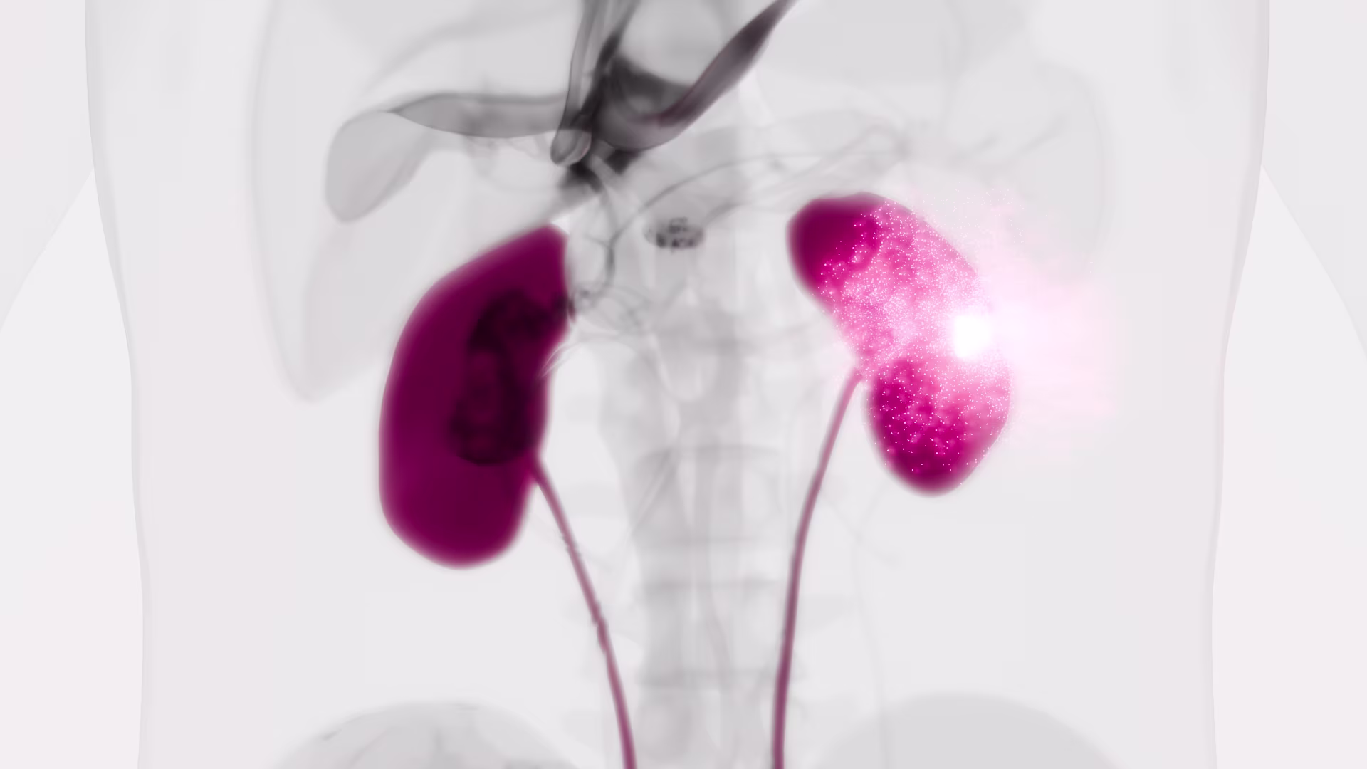
Background: Single circulating tumor cells (CTCs) or circulating tumor microemboli (CTMs) are potential biomarkers of renal cell cancer (RCC), however studies of CTCs/CTMs in RCC are limited. In this pilot study we aimed to evaluate a novel blood filtration technique suited for cytomorphological classification, immunocytochemical and molecular characterization of filtered, so called circulating non-hematologic cells (CNHCs) – putative CTCs/CTMs – in patients with RCC.
Methods: Blood of 40 patients with renal tumors was subjected to ScreenCell filtration. CNHCs were classified according to cytomorphological criteria. Immunocytochemical analysis was performed with antibodies against CD45, CD31 and carbonic anhydrase IX (CAIX, a RCC marker). DNA of selected CNHCs and respective primary tumors was analysed by array-CGH.
Results: CNHC-clusters with malignant or uncertain malignant cytomorphological features – putative CTMs – were negative for CD45, positive for CD31, while only 6% were CAIX positive. Array-CGH revealed that 83% of malignant and uncertain malignant cells did represent with a balanced genome whereas 17% presented genomic DNA imbalances which did not match the aberrations of the primary tumors. Putative single CTCs were negative for CD45, 33% were positive for CD31 and 56% were positive for CAIX.
Conclusions: The majority of CNHC-clusters, putative CTMs, retrieved by ScreenCell filtration may be of endothelial origin. Morphological criteria seem to be insufficient to distinguish malignant from non-malignant cells in renal cancer.
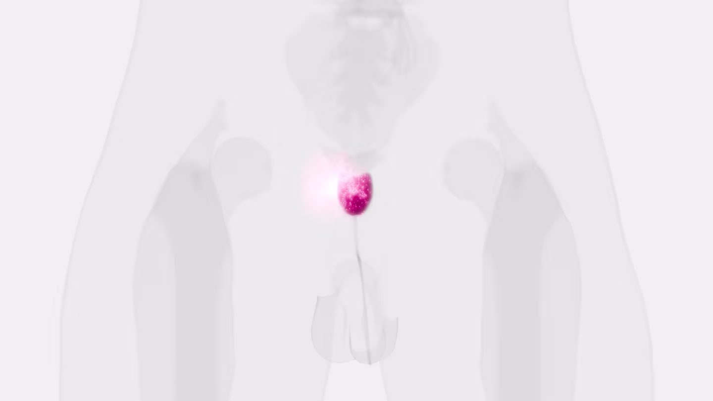
Detection of circulating tumor cells in patients with adrenocortical carcinoma: a monocentric preliminary study
Context: Adrenocortical carcinoma (ACC) is a rare malignancy, the prognosis of which is mainly dependent on stage at diagnosis. The identification of disease-associated markers for early diagnosis and drug monitoring is mandatory. Circulating tumor cells (CTCs) are released into the bloodstream from primary tumor/metastasis. CTC detection in blood samples may have enormous potential for assisting in the diagnosis of malignancy, estimating prognosis, and monitoring the disease.
Objective: The aim of the study was to investigate the presence of CTCs in blood samples of patients with ACC or benign adrenocortical adenoma (ACA).
Setting: We conducted the study at a university hospital.
Intervention: CTC analysis was performed in blood samples from 14 ACC patients and 10 ACA patients. CTCs were isolated on the basis of cell size by filtration through ScreenCell devices, followed by identification according to validated morphometric criteria and immunocytochemistry.
Main outcome measure: We measured the difference in CTC detection between ACC and ACA.
Results: CTCs were detected in all ACC samples, but not in ACA samples. Immunocytochemistry confirmed the adrenocortical origin. When ACC patients were stratified according to the median value of tumor diameter and metastatic condition, a statistically significant difference was found in the number of CTCs detected after surgery. A significant correlation between the number of CTCs in postsurgical samples and clinical parameters was found for tumor diameter alone.
Conclusions: Our findings provide the first evidence for adrenocortical tumors that CTCs may represent a useful marker to support differential diagnosis between ACC and ACA. The correlation with some clinical parameters suggests a possible relevance of CTC analysis for prognosis and noninvasive monitoring of disease progression and drug response.
A new device for rapid isolation by size and characterization of rare circulating tumor cells
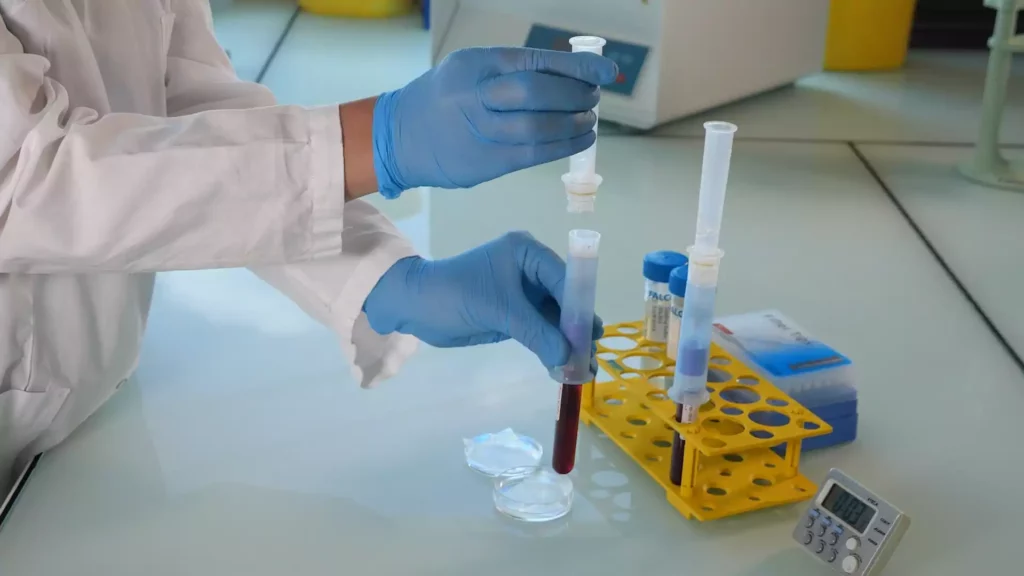
Background: Circulating tumor cells (CTCs) likely derive from clones in the primary tumor, suggesting that they can be used for all biological tests applying to the primary cells.
Materials and methods: The ScreenCell® devices are single-use and low-cost innovative devices that use a filter to isolate and sort tumor cells by size.
Results: The ScreenCell® Cyto device is able to isolate rare, fixed, tumor cells, with a high recovery rate. Cells are well preserved morphologically. Immunocytochemistry and FISH assays can be performed directly on the filter. The ScreenCell® CC device allows isolation of live cells able to grow in culture. High quality genetic materials can be obtained directly from tumor cells isolated on the ScreenCell® MB device filter.
Conclusion: Due to their reduced size, versatility, and capacity to isolate CTCs within minutes, the ScreenCell® devices may be able to simplify and improve non-invasive access to tumor cells.