
Non-Metastatic Esophageal Adenocarcinoma: Circulating Tumor Cells in the Course of Multimodal Tumor Treatment
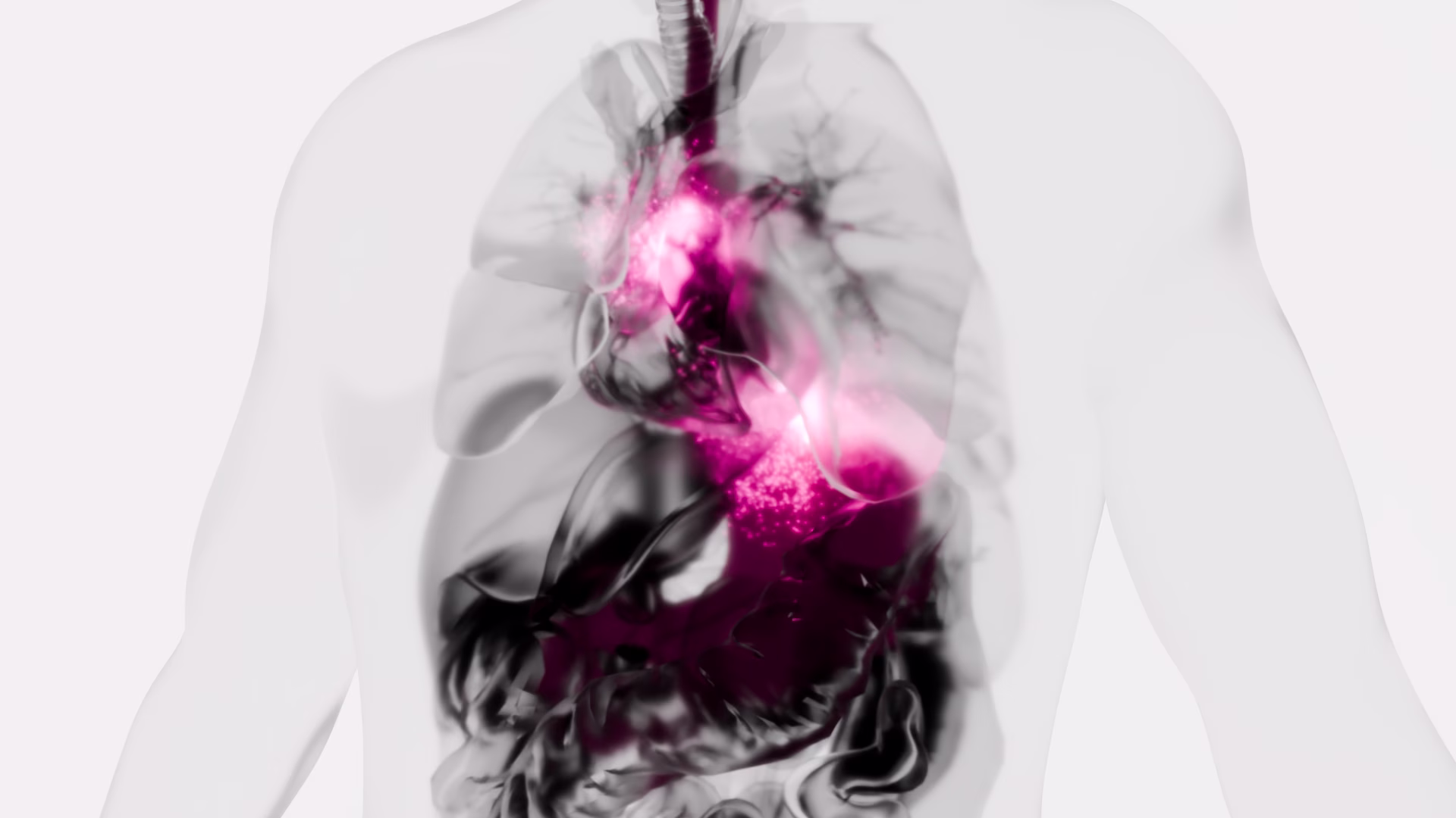
Background: Isolation of circulating tumor cells (CTC) holds the promise to improve response-prediction and personalization of cancer treatment. In this study, we test a filtration device for CTC isolation in patients with non-metastatic esophageal adenocarcinoma (EAC) within recent multimodal treatment protocols. Methods: Peripheral blood specimens were drawn from EAC patients before and after neoadjuvant chemotherapy (FLOT)/chemoradiation (CROSS) as well as after surgery. Filtration using ScreenCell® devices captured CTC for cytologic analysis. Giemsa-stained specimens were evaluated by a cytopathologist; the cut-off was 1 CTC/specimen (6 mL). Immunohistochemistry with epithelial (pan-CK) and mesenchymal markers (vimentin) was performed. Results: Morphologically diverse malignant CTCs were found in 12/20 patients in at least one blood specimen. CTCs were positive for both vimentin and pan-CK. More patients were CTC positive after neoadjuvant therapy (6/20 vs. 9/15) and CTCs per/ml increased in most of the CTC-positive patients. After surgery, 8/13 patients with available blood specimens were still CTC positive. In clinical follow-up, 5/9 patients who died were CTC-positive. Conclusions: Detection of CTC by filtration within multimodal treatment protocols of non-metastatic EAC is feasible. The rate of CTC positive findings and the quantity of CTCs changes in the course of multimodal neoadjuvant chemoradiation/chemotherapy and surgery.
Isolation and characterization of circulating melanoma cells by size filtration and fluorescent in-situ hybridization.

Isolation of circulating tumor cells (CTCs) from blood of melanoma patients has been difficult owing to inconsistent expression of surface antigens. Here we report on the isolation, detection, and characterization of CTCs from blood of melanoma patients using microfiltration and fluorescent in-situ hybridization (FISH). Two tubes of blood from 15 patients with advanced melanoma were collected. These two tubes subsequently underwent filtration through a membrane with pore sizes of 7.5 μm. Isolated cells from one tube were analyzed by FISH for RREB1 (6p24), MYB (6q32), SE6 (D6Z1), and CCND1 (11q13) and the other paired specimen was analyzed by immunofluorescence for HMB45, melanoma-associated antigen recognized by T cells-1, tyrosinase and melanogenesis associated transcription factor. We identified CTCs in 10 out of 13 melanoma samples by immunofluorescence (2.5–99 CTCs/3 ml of blood) and in 13 specimens by FISH (7.2–76 CTCs/3 ml of blood) with more CTCs identified by FISH in 10 out 13 samples. Two filters failed. Our results show that CTCs are detectable in the majority of patients with advanced melanoma. These tools will be useful in characterizing treatment related changes of melanoma in CTCs.
Liquid Biopsy in Rare Cancers: Lessons from Hemangiopericytoma.
Hemangiopericytoma (HPT) is a rare mesenchymal tumor of fibroblastic type and for its rarity is poorly studied. The most common sites of metastatic disease in patients with intracranial HPT are the bone, liver, and lung, suggestive for an hematogenous dissemination; for this reason, we investigated, for the first time, the presence of circulating tumor cells (CTCs) in hemangiopericytoma patient by CellSearch® and SceenCell® devices. Peripheral blood samples were drawn and processed by CellSearch, an EpCAM-dependent device, and ScreenCell®, a device size based. We found nontypical CTCs by CellSearch system and the immunofluorescence analysis performed on CTCs isolate by ScreenCell demonstrated the presence of single CTCs and CTC clusters. The molecular characterization of single CTCs and CTC clusters, using antibodies directed against EpCAM, CD34, cytokeratins (8, 18, and 19), and CD45, showed a great heterogeneity in CTC clusters. We believe that the present study may open a new scenario in the rare tumors: the introduction of the liquid biopsy and the molecular characterization of circulating tumor cells could lead to personalized targeted treatments and also for rare tumors.
Circulating Tumor DNA Reflects Tumor Metabolism Rather Than Tumor Burden in Chemotherapy-Naive Patients with Advanced Non-Small Cell Lung Cancer: 18F-FDG PET/CT Study

We aimed to evaluate the relationships between circulating tumor cells (CTCs) or plasma cell–free DNA (cfDNA) on one side and a comprehensive range of 18F-FDG PET/CT–derived parameters on the other side in chemotherapy-naive patients with advanced non–small cell lung cancer (NSCLC). Methods: From a group of 79 patients included in a trial evaluating the role of pretreatment circulating tumor markers as predictors of prognosis in chemotherapy-naive patients with advanced NSCLC, we recruited all those who underwent 18F-FDG PET/CT for clinical reasons at our institution before inclusion in the trial (and thus just before chemotherapy). For each patient, a peripheral blood sample was collected at baseline for the evaluation of CTCs and cfDNA. CTCs were isolated by size using a filtration-based device and then morphologically identified and enumerated; cfDNA was isolated from plasma and quantified by a quantitative polymerase chain reaction using human telomerase reverse transcriptase. The following 18F-FDG PET/CT–derived parameters were computed: maximum diameter of the primary lesion (T), of the largest lymph node (N), and of the largest metastatic lesion (M); SUVmax; SUVmean; size-incorporated SUVmax; metabolic tumor volume; and total lesion glycolysis. All parameters were independently measured for T, N, and M. The associations among CTCs, cfDNA, and 18F-FDG PET/CT–derived parameters were evaluated by multivariate-analysis. Patients were divided into 2 groups according to the presence of either limited metastatic involvement (M1a or M1b due to extrathoracic lymph nodes only) or disseminated metastatic disease. The presence or absence of metabolically active bone lesions was also recorded for each patient, and patient subgroups were compared. Results: Thirty-seven patients recruited in the trial matched our PET-based criteria (24 men; age, 64.5 ± 8.1 y). SUVmax for the largest metastatic lesion was the only variable independently associated with baseline cfDNA levels (P = 0.016). Higher levels of cfDNA were detected in the subgroup of patients with metabolically active bone lesions (P = 0.02), but no difference was highlighted when patients with more limited metastatic disease were compared with patients with disseminated metastatic disease. Conclusion: The correlation of cfDNA levels with tumor metabolism, but not with metabolic tumor volume at regional or distant levels, suggests that cfDNA may better reflect tumor biologic behavior or aggressiveness rather than tumor burden in metastatic NSCLC.
EpCAM-expressing circulating tumor cells in colorectal cancer

Background: Several studies have raised the issue of the inadequacy of CellSearch® to detect the entire pool of circulating tumor cells (CTCs) from blood of cancer patients, suggesting that cells expressing low levels of epithelial cell adhesion molecule (EpCAM) are not recognized by the capture reagent. In this exploratory study, we aimed to evaluate the status of EpCAM in CTCs isolated from a group of metastatic colorectal cancer patients, in 40% of whom, CTC had been found to be undetected by the CellSearch® system.
Methods: CTCs were analyzed using both a microfiltration method (ScreenCell) and CellSearch® in parallel. Furthermore, since EpCAM exists in 2 different variants, we investigated the presence of both its intracellular domain (EpICD) and extracellular domain (EpEX) through immunofluorescence staining of CTCs on filters.
Results: Results from immunofluorescence experiments demonstrated that, overall, EpICD and/or EpEX was expressed in 176 CTCs detected by ScreenCell, while the CellSearch® system was able to capture only 10 CTCs.
Conclusions: This is the first demonstration that the low sensitivity of CellSearch® to detect CTCs in colorectal cancer patients is not due to the lack of EpCAM. »
Rapid and Sensitive Detection of Breast Cancer Cells in Patient Blood with Nuclease-Activated Probe Technology

A challenge for circulating tumor cell (CTC)-based diagnostics is the development of simple and inexpensive methods that reliably detect the diverse cells that make up CTCs. CTC-derived nucleases are one category of proteins that could be exploited to meet this challenge. Advantages of nucleases as CTC biomarkers include: (1) their elevated expression in many cancer cells, including cells implicated in metastasis that have undergone epithelial-to-mesenchymal transition; and (2) their enzymatic activity, which can be exploited for signal amplification in detection methods. Here, we describe a diagnostic assay based on quenched fluorescent nucleic acid probes that detect breast cancer CTCs via their nuclease activity. This assay exhibited robust performance in distinguishing breast cancer patients from healthy controls, and it is rapid, inexpensive, and easy to implement in most clinical labs. Given its broad applicability, this technology has the potential to have a substantive impact on the diagnosis and treatment of many cancers.
Perioperative detection of circulating tumour cells in patients with lung cancer.
Lung cancer is a leading cause of mortality and despite surgical resection a proportion of patients may develop metastatic spread. The detection of circulating tumour cells (CTCs) may allow for improved prediction of metastatic spread and survival. The current study evaluates the efficacy of the ScreenCell® filtration device, to capture, isolate and propagate CTCs in patients with primary lung cancer. Prior to assessment of CTCs, the present study detected cancer cells in a proof‑of‑principle‑ experiment using A549 human lung carcinoma cells as a model. Ten patients (five males and five females) with pathologically diagnosed primary non‑small cell lung cancer undergoing surgical resection, had their blood tested for CTCs. Samples were taken from a peripheral vessel at the baseline, from the pulmonary vein draining the lobe containing the tumour immediately prior to division, a further central sample was taken following completion of the resection, and a final peripheral sample was taken three days post‑resection. A significant increase in CTCs was observed from baseline levels following lung manipulation. No association was able to be made between increased levels of circulating tumour cells and survival or the development of metastatic deposits. Manipulation of the lung during surgical resection for non‑small cell lung carcinoma results in a temporarily increased level of CTCs; however, no clinical impact for this increase was observed. Overall, the study suggests the ScreenCell® device has the potential to be used as a CTC isolation tool, following further work, adaptations and improvements to the technology and validation of results.
Circulating Cell-Free DNA and Circulating Tumor Cells as Prognostic and Predictive Biomarkers in Advanced Non-Small Cell Lung Cancer Patients Treated with First-Line Chemotherapy
Cell-free DNA (cfDNA) and circulating tumor cells (CTCs) are promising prognostic and predictive biomarkers in non-small cell lung cancer (NSCLC). In this study, we examined the prognostic role of cfDNA and CTCs, in separate and joint analyses, in NSCLC patients receiving first line chemotherapy. Seventy-three patients with advanced NSCLC were enrolled in this study. CfDNA and CTC were analyzed at baseline and after two cycles of chemotherapy. Plasma cfDNA quantification was performed by quantitative PCR (qPCR) whereas CTCs were isolated by the ScreenCell Cyto (ScreenCell, Paris, France) device and enumerated according to malignant features. Patients with baseline cfDNA higher than the median value (96.3 hTERT copy number) had a significantly worse overall survival (OS) and double the risk of death (hazard ratio (HR): 2.14; 95% confidence limits (CL) = 1.24–3.68; p-value = 0.006). Conversely, an inverse relationship between CTC median baseline number (6 CTC/3 mL of blood) and OS was observed. In addition, we found that in patients reporting stable disease (SD), the baseline cfDNA and CTCs were able to discriminate patients at high risk of poor survival. cfDNA demonstrated a more reliable biomarker than CTCs in the overall population. In the subgroup of SD patients, both biomarkers identified patients at high risk of poor prognosis who might deserve additional/alternative therapeutic interventions.
Cytologic Characteristics of Circulating Epithelioid Cells in Pancreatic Disease
BACKGROUND
Circulating epithelioid cells (CECs), also known as circulating tumor, circulating cancer, circulating epithelial, or circulating nonhematologic cells, are a prognostic factor in various malignancies that can be isolated via various protocols. In the current study, the authors analyzed the cytomorphologic characteristics of CECs isolated by size in a cohort of patients with benign and malignant pancreatic diseases to determine whether cytomorphological features could predict CEC origin.
METHODS
Blood samples were collected from 9 healthy controls and 171 patients with pancreatic disease who were presenting for surgical evaluation before treatment. Blood was processed with the ScreenCell size-based filtration device. Evaluable CECs were analyzed in a blinded fashion for cytomorphologic characteristics, including cellularity; nucleoli; nuclear size, irregularity, variability, and hyperchromasia; and nuclear-to-cytoplasmic ratio. Statistical differences between variables were analyzed via the Fisher exact test.
RESULTS
No CECs were identified among the 9 normal healthy controls. Of the 115 patients with CECs (positive or suspicious for), 25 had nonmalignant disease and 90 had malignancy. There were no significant differences in any of the cytologic criteria noted between groups divided by benign versus malignant, neoplastic versus nonneoplastic, or pancreatic ductal adenocarcinoma versus neuroendocrine tumor.
CONCLUSIONS
CECs were observed in patients with malignant and nonmalignant pancreatic disease, but not in healthy controls. There were no morphologic differences observed between cells from different pancreatic diseases, suggesting that numerous conditions may be associated with CECs in the circulation and that care must be taken not to overinterpret cells identified by cytomorphology as indicative of circulating tumor cells of pancreatic cancer. Additional studies are required to determine the origin and clinical significance of these cells. »
Detection of Circulating Tumour Cells in Urothelial Cancers and Clinical Correlations: Comparison of Two Methods
Circulating tumour cells (CTC) are identified exploiting their protein/gene expression patterns or distinct size compared to blood cells. Data on CTC in bladder cancer (BC) are still scarce. We comparatively analyzed CTC enrichment by AdnaTest ProstateCancerSelect (AT) and ScreenCell®Cyto (SC) kits, combined with identification by EPCAM, MUC1, and ERBB2 expression and by cytological criteria, respectively, in 19 nonmetastatic (M0) and 47 metastatic (M+) BC patients, at baseline (T0) and during treatment (T1). At T0, CTC positivity rates by AT were higher in M+ compared to M0 cases (57.4% versus 25%, p = 0.041). EPCAM was detected in 75% of CTC-positive samples by AT, showing increasing expression levels from T0 to T1 (median (interquartile range, IQR): 0.18 (0.07–0.42) versus 0.84 (0.33–1.84), p = 0.005) in M+ cases. Overall, CTC positivity by SC was around 80% regardless of clinical setting and time point of analysis, except for a lower occurrence at T1 in M0 cases. At T0, circulating tumour microemboli were more frequently (25% versus 8%) detected and more numerous in M+ compared to M0 patients. The approach used for CTC detection impacts the outcome of CTC studies. Further investigations are required to clarify the clinical validity of AT and SC in specific BC clinical contexts.