
An assessment of diagnostic performance of a filter-based antibody-independent peripheral blood circulating tumour cell capture paired with cytomorphologic criteria for the diagnosis of cancer
Objectives
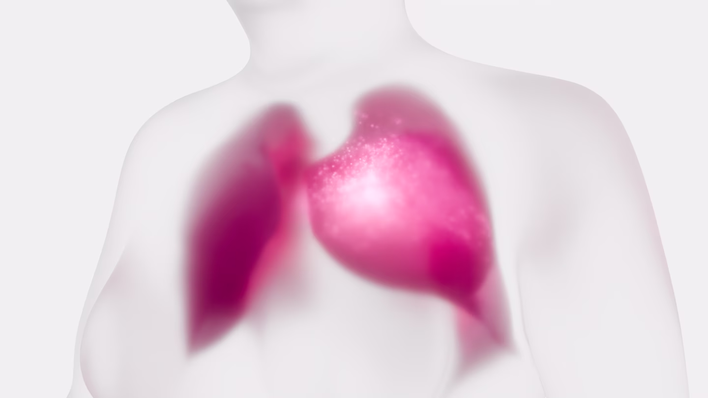
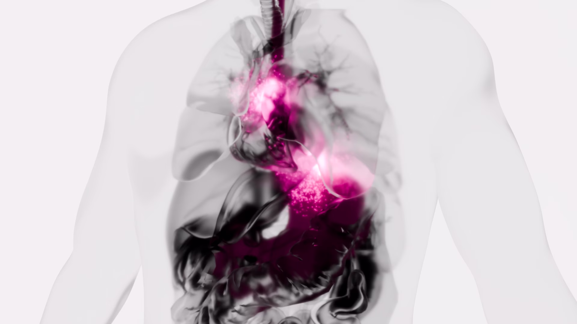
Circulating tumour cells (CTCs) are reported to be predictive for prognosis and response to treatment in advanced lung cancer. However, the clinical utility of the CTCs detection remains unknown for early stage lung cancer as the number of CTCs is reported as low, providing challenges in identification. We have evaluated diagnostic performance of filtration-based technology using cytomorphologic criteria in patients undergoing surgery for lung cancer.
Material and methods
We processed blood from 76 patients undergoing surgery for known or suspected lung cancer using ScreenCell® Cyto filter devices. Captured cells were stained using haematoxylin and eosin and independently assessed by two pathologists for the presence of atypical cells suspicious for cancer. Diagnostic performance was evaluated against pathologist reported diagnoses of cancer from surgically obtained specimens.
Results
Cancer was diagnosed in 57 patients (77.0%), including 32 with primary lung cancer (56.1%). The proportion of patients with early stage primary lung cancer in which CTCs were identified was 18 and 21 (56.3% and 65.6%, respectively) as reported by two pathologists. The agreement between the pathologists was 77.0% corresponding to a kappa-statistic of 53.7% indicating moderate agreement. No significant differences were found for the percentage of CTCs for primary and metastatic cancer as well as for cancer stages. On sensitivity weighted analysis, a sensitivity and specificity were 71.9% (95% CI 60.5–83.0) and 52.9% (95% CI 31.1–77.0), respectively. On specificity weighted analysis, a sensitivity and specificity were 50.9% (95% CI 39.3–64.4) and 82.4% (60.4–96.2), respectively.
Conclusion
The performance of the tested filter-based antibody-independent technology to capture CTCs using standard cytomorphologic criteria provides the potential of a diagnostic blood test for lung cancer.
Cancer-associated Macrophage-like Cells in Patients with Non-metastatic Adenocarcinoma of the Esophagus – Cytomorphological Heterogeneity
Introduction: Esophageal adenocarcinoma (EAC) often recurs systemically despite therapy with a curative aim. New diagnostic and therapeutic approaches are urgently needed. A promising field is liquid biopsy, meaning the investigation of tumor-associated cells in the peripheral blood, for example cancer-associated macrophage-like cells (CAML). The aim of this multicentric study was to investigate the presence and cytomorphological appearance of CAML in patients with non-metastatic and operable esophageal cancer.
Methods: Blood samples from 252 patients with locally advanced EAC were obtained before starting curative treatment including surgery, and then processed using ScreenCell® filtration devices. Cytological analysis was performed via May-Grünwald-Giemsa staining. CAML were defined by their morphological characteristics. We also performed immunofluorescence staining with the mesenchymal marker vimentin on a subset of our study cohort.
Results: We detected cytomorphologically heterogeneous CAML in 31.8% (n=80) patients. Their presence and cell count did not correlate significantly with pretherapeutic cTNM. Even in patients with small tumors and no lymph-node infiltration, cell counts were high. CAML showed heterogenous staining patterns for vimentin.
Conclusion: This is one of the first studies demonstrating the presence and phenotype of CAML in a uniquely broad cohort of EAC patients. As they are believed to be representatives of the inflammatory tumor microenvironment shed into the bloodstream, their presence in non-metastatic EAC is a promising finding.
Cytomorphologic visualization of circulating tumor cells in urinary bladder cancer patients using ScreenCell™ technology: Potential as a simple cytology test

Circulating tumor cells (CTC) are a recent technique which is a potentially important prognostic factor in many solid tumors. There are many techniques of detecting CTCs, but they usually implement costly techniques like EpCAM targeted detection, fluorescence-based diagnosis, or magnetic bead based positive or negative selection. The diagnostic utility of simple cytomorphological diagnosis after routine staining of CTCs have been rarely studied. We aimed to detect CTCs in 24 patients clinically suspected to have Urinary Bladder Cancer using a simple but efficient patented filtration technology (ScreenCell™), followed by optical microscopic visualization after routine May-Grunwald-Giemsa (MGG) staining. The detected CTCs were then tested for association with the histologic type, lamina propria invasion, deep muscle invasion and the T-stage. Out of the 24 patients tested, one was found to have papilloma, nine had low grade urothelial carcinoma, 13 had high grade urothelial carcinoma and one had poorly differentiated adenocarcinoma. Of these, two LGUC, eight HGUC and one adenocarcinoma had detectable CTC. Presence of CTCs had a statistically significant association with Lamina propria invasion (P = .006) and T-stage (P = .02), and a trend toward significance for differentiating LGUC from HGUC (P = .10). These results suggest that cytomorphological detection of CTC is likely to be clinically useful in diagnosis and prognostication of urinary blader cancers. These findings need to be confirmed on studies with larger sample sizes.
Gene signatures of circulating breast cancer cell models are a source of novel molecular determinants of metastasis and improve circulating tumor cell detection in patients

Background: Progression to stage IV disease remains the main cause of breast cancer-related deaths. Increasing knowledge on the hematogenous phase of metastasis is key for exploiting the entire window of opportunity to interfere with early dissemination and to achieve a more effective disease control. Recent evidence suggests that circulating tumor cells (CTCs) possess diverse adaptive mechanisms to survive in blood and eventually metastasize, encouraging research into CTC-directed therapies.
Methods: On the hypothesis that the distinguishing molecular features of CTCs reveal useful information on metastasis biology and disease outcome, we compared the transcriptome of CTCs, primary tumors, lymph-node and lung metastases of the MDA-MB-231 xenograft model, and assessed the biological role of a panel of selected genes, by in vitro and in vivo functional assays, and their clinical significance in M0 and M+ breast cancer patients.
Results: We found that hematogenous dissemination is governed by a transcriptional program and identified a CTC signature that includes 192 up-regulated genes, mainly related to cell plasticity and adaptation, and 282 down-regulated genes, involved in chromatin remodeling and transcription. Among genes up-regulated in CTCs, FADS3 was found to increases cell membrane fluidity and promote hematogenous diffusion and lung metastasis formation. TFF3 was observed to be associated with a subset of CTCs with epithelial-like features in the experimental model and in a cohort of 44 breast cancer patients, and to play a role in cell migration, invasion and blood-borne dissemination. The analysis of clinical samples with a panel of CTC-specific genes (ADPRHL1, ELF3, FCF1, TFF1 and TFF3) considerably improved CTC detection as compared with epithelial and tumor-associated markers both in M0 and stage IV patients, and CTC kinetics informed disease relapse in the neoadjuvant setting.
Conclusions: Our findings provide evidence on the potential of a CTC-specific molecular profile as source of metastasis-relevant genes in breast cancer experimental models and in patients. Thanks to transcriptome analysis we generated a novel CTC signature in the MDA-MB-231 xenograft model, adding a new piece to the current knowledge on the key players that orchestrate tumor cell hematogenous dissemination and breast cancer metastasis, and expanding the list of CTC-related biomarkers for future validation studies.
Strategies for Isolating and Propagating Circulating Tumor Cells in Men with Metastatic Prostate Cancer
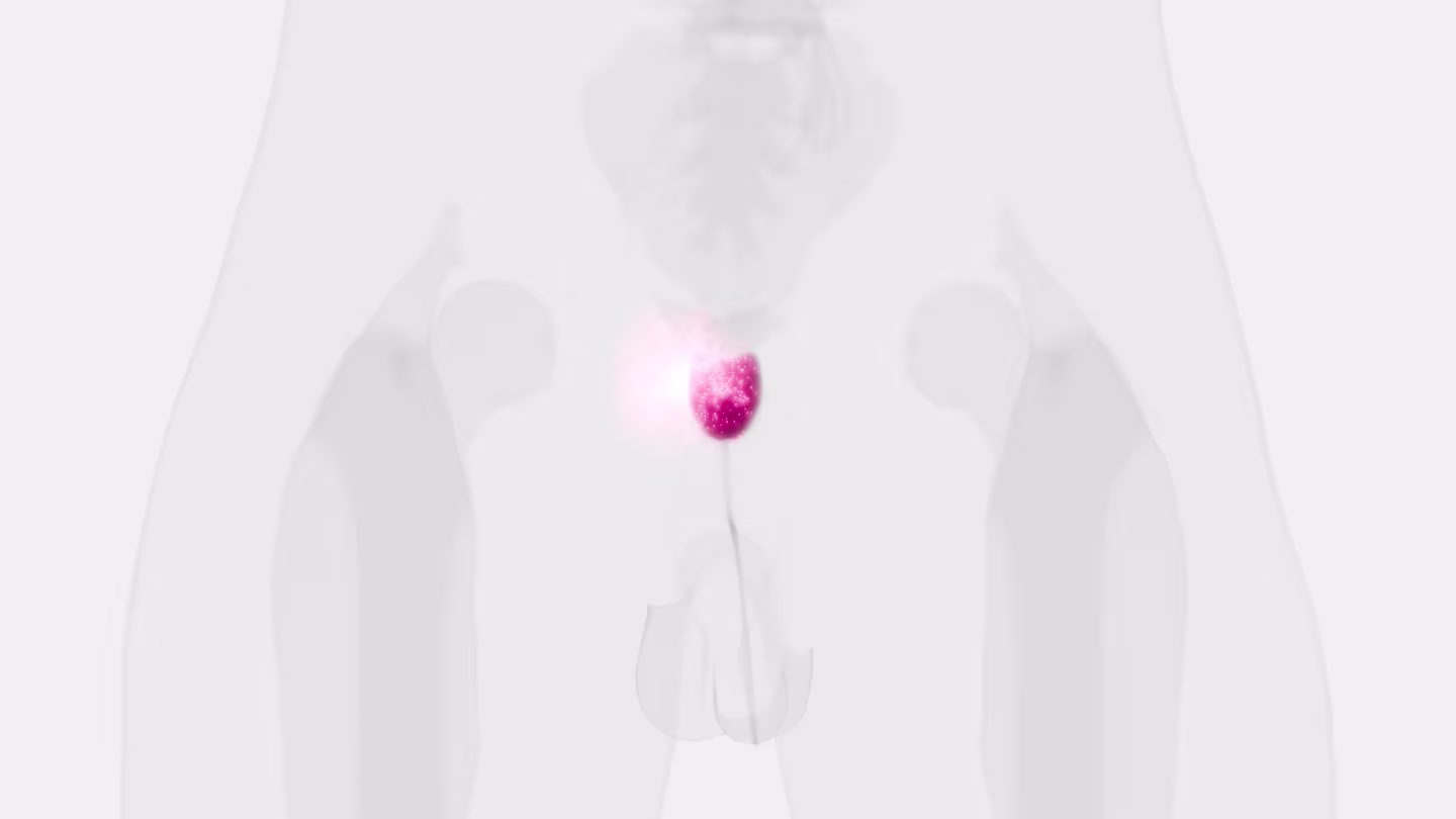
Selecting a well-suited method for isolating/characterizing circulating tumor cells (CTCs) is challenging. Evaluating sensitive and specific markers for prostate cancer (PCa)-specific CTC identification and analysis is crucial. We used the CellCollector EpCAM-functionalized system (CC-EpCAM) and evaluated and developed a PCa-functionalized version (CC-PCa); we then compared CTC isolation techniques that exploit the physical and biological properties of CTCs. We established two cohorts of metastatic PCa patients (mPCa; 15 in cohort 1 and 10 in cohort 2). CTC cultivation experiments were conducted with two capturing methods (Ficoll and ScreenCell). The most sensitive detection rates and highest CTC counts were reached with the CC-PCa and ScreenCell system. Patients with ≥5 CTCs isolated with CC-EpCAM had an overall survival (OS) of 0.93 years, and patients with ≥5 CTCs isolated with CC-PCa had an OS of 1.5 years in cohort 1. Nevertheless, we observed the highest sensitivity and specificity for 24-month survival by the Ficoll with CD45 depletion and ScreenCell system with May-Grunwald Giemsa (MGG) staining. The EpCAM molecule is an essential factor related to OS for CTC isolation based on biological properties in mPCa patients. The best-suited CTC capture system is not limited to one characteristic of cells but adapted to downstream analysis.
Identification of Atypical Circulating Tumor Cells with Prognostic Value in Metastatic Breast Cancer Patients
Circulating tumor cells have a strong potential as a quasi-non-invasive tool for setting up a precision medicine strategy for cancer patients. Using a second-generation « »filtration-based » » technology to isolate CTCs, the Screencell™ technology (Sarcelles, France), we performed a large and simultaneous analysis of all atypical circulating tumor cells (aCTCs) isolated from the blood of metastatic breast cancer (mBC) patients. We correlated their presence with clinicopathological and survival data. We included 91 mBC patients from the PERMED-01 study. The median number of aCTCs was 8.3 per mL of blood. Three subsets of aCTCs, absent from controls, were observed in patients: single (s-aCTCs), circulating tumor micro-emboli (CTM), and giant-aCTCs (g-aCTCs). The presence of g-aCTCs was associated with shorter progression free survival and overall survival. This study highlights the heterogeneity of aCTCs in mBC patients both at the cytomorphological and molecular levels. In addition, it suggests the usefulness of the g-aCTC subset as a prognostic factor and a potential stratification tool to treat late-stage mBC patients and improve their chances of benefiting from early clinical trials.
Sequential Isolation and Characterization of Single CTCs and Large CTC Clusters in Metastatic Colorectal Cancer Patients
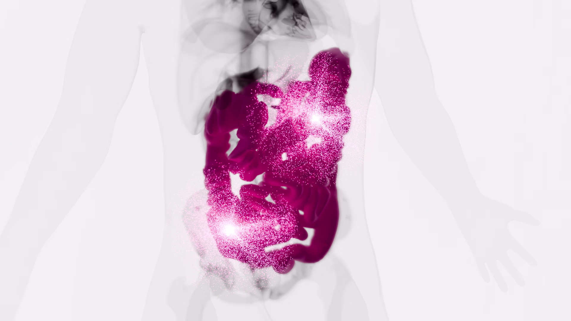
Circulating tumor cells (CTCs) detach from a primary tumor or its metastases and circulate in the bloodstream. The vast majority of CTCs are deemed to die into the bloodstream, with only few cells representing viable metastatic precursors. Particularly, single epithelial CTCs do not survive long in the circulation due to the loss of adhesion-dependent survival signals. In metastatic colorectal cancer, the generation of large CTC clusters is a very frequent occurrence, able to increase the aptitude of CTCs to survive in the bloodstream. Although a deepened analysis of large-sized CTC clusters might certainly offer new insights into the complexity of the metastatic cascade, most CTC isolation techniques are unfortunately not compatible with large-sized CTC clusters isolation. The inappropriateness of standard CTC isolation devices for large clusters isolation and the scarce availability of detection methods able to specifically isolate and characterize both single CTCs and CTC clusters finally prevented in-depth studies on the prognostic and predictive value of clusters in clinical practice, unlike that which has been described for single CTCs. In the present study, we validated a new sequential filtration method for the simultaneous isolation of large CTC clusters and single CTCs in patients with metastatic colorectal cancer at failure of first-line treatments. The new method might allow differential downstream analyses for single and clustered CTCs starting from a single blood draw, opening new scenarios for an ever more precise characterization of colorectal cancer metastatic cascade.
Circulating Tumor Cell Clusters Are Frequently Detected in Women with Early-Stage Breast Cancer
The clinical relevance of circulating tumor cell clusters (CTC-clusters) in breast cancer (BC) has been mostly studied using the CellSearch®, a marker-dependent method detecting only epithelial-enriched clusters. However, due to epithelial-to-mesenchymal transition, resorting to marker-independent approaches can improve CTC-cluster detection. Blood samples collected from healthy donors and spiked-in with tumor mammospheres, or from BC patients, were processed for CTC-cluster detection with 3 technologies: CellSearch®, CellSieve™ filters, and ScreenCell® filters. In spiked-in samples, the 3 technologies showed similar recovery capability, whereas, in 19 clinical samples processed in parallel with CellSearch® and CellSieve™ filters, filtration allowed us to detect more CTC-clusters than CellSearch® (median number = 7 versus 1, p = 0.0038). Next, samples from 37 early BC (EBC) and 23 metastatic BC (MBC) patients were processed using ScreenCell® filters for attaining both unbiased enrichment and marker-independent identification (based on cytomorphological criteria). At baseline, CTC-clusters were detected in 70% of EBC cases and in 20% of MBC patients (median number = 2, range 0-20, versus 0, range 0-15, p = 0.0015). Marker-independent approaches for CTC-cluster assessment improve detection and show that CTC-clusters are more frequent in EBC than in MBC patients, a novel finding suggesting that dissemination of CTC-clusters is an early event in BC natural history.
Cytopathological Heterogeneity of Circulating Tumor Cells in Non-metastatic Esophageal Adenocarcinoma
Background/Aim: The presence of circulating tumor cells (CTC) has been reported to have an impact on prognosis in different tumor entities. Little is known about CTC morphology and heterogeneity. Patients and Methods: In a multicenter setting, pre-therapeutic peripheral blood specimens were drawn from patients with non-metastatic esophageal adenocarcinoma (EAC). CTCs were captured by size-based filtration (ScreenCell®), subsequently Giemsa-stained and evaluated by two trained readers. The isolated cells were categorized in groups based on morphologic criteria. Results: Small and large single CTCs, as well as CTC-clusters, were observed in 69.2% (n=81) of the 117 specimens; small CTCs were observed most frequently (59%; n=69), followed by large CTCs (40%; n=47) and circulating cancer-associated macrophage-like cells (CAMLs; 34.2%, n=40). Clusters were rather rare (12%; n=14). CTC/CAML were heterogeneous in the cohort, but also within one specimen. Neither the presence of the CTC subtypes/CAMLs nor the exact cell count were associated with the primary clinical TNM stage. Conclusion: Morphologically heterogenic CTCs and CAMLs are present in patients with non-metastatic, non-pretreated EAC.
Isolation and Enumeration of CTC in Colorectal Cancer Patients: Introduction of a Novel Cell Imaging Approach and Comparison to Cellular and Molecular Detection Techniques
Circulating tumour cells (CTC) were proven to be prognostically relevant in cancer treatment, e.g., in colorectal cancer (CRC). This study validates a molecular detection technique through using a novel cell imaging approach for CTC detection and enumeration, in comparison to a size-based cellular and correlated the data to clinico-pathological characteristics. Overall, 57 CRC patients were recruited for this prospective study. Blood samples were analysed for CTCs by three methods: (1) Epithelial marker immunofluorescence staining combined with automated microscopy using the NYONE® cell imager; (2) isolation by size using membrane filtration with the ScreenCell® Cyto IS device and immunofluorescence staining; (3) detection by semi-quantitative Cytokeratin-20 RT-qPCR. Enumeration data were compared and correlated with clinic-pathological parameters. CTC were detected by either approach; however, with varying positivity rates: NYONE® 36.4%, ScreenCell® 100%, and PCR 80.5%. All methods revealed a positive correlation of CTC presence and higher tumour burden, which was most striking using the ScreenCell® device. Generally, no intercorrelation of CTC presence emerged amongst the applied techniques. Overall, enumeration of CTC after isolation by size demonstrated to be the most reliable strategy for the detection of CTC in CRC patients. Ongoing studies will have to unravel the prognostic value of this finding, and validate this approach in a larger cohort.