
Liquid Biopsy in Rare Cancers: Lessons from Hemangiopericytoma.
Hemangiopericytoma (HPT) is a rare mesenchymal tumor of fibroblastic type and for its rarity is poorly studied. The most common sites of metastatic disease in patients with intracranial HPT are the bone, liver, and lung, suggestive for an hematogenous dissemination; for this reason, we investigated, for the first time, the presence of circulating tumor cells (CTCs) in hemangiopericytoma patient by CellSearch® and SceenCell® devices. Peripheral blood samples were drawn and processed by CellSearch, an EpCAM-dependent device, and ScreenCell®, a device size based. We found nontypical CTCs by CellSearch system and the immunofluorescence analysis performed on CTCs isolate by ScreenCell demonstrated the presence of single CTCs and CTC clusters. The molecular characterization of single CTCs and CTC clusters, using antibodies directed against EpCAM, CD34, cytokeratins (8, 18, and 19), and CD45, showed a great heterogeneity in CTC clusters. We believe that the present study may open a new scenario in the rare tumors: the introduction of the liquid biopsy and the molecular characterization of circulating tumor cells could lead to personalized targeted treatments and also for rare tumors.
EpCAM-expressing circulating tumor cells in colorectal cancer

Background: Several studies have raised the issue of the inadequacy of CellSearch® to detect the entire pool of circulating tumor cells (CTCs) from blood of cancer patients, suggesting that cells expressing low levels of epithelial cell adhesion molecule (EpCAM) are not recognized by the capture reagent. In this exploratory study, we aimed to evaluate the status of EpCAM in CTCs isolated from a group of metastatic colorectal cancer patients, in 40% of whom, CTC had been found to be undetected by the CellSearch® system.
Methods: CTCs were analyzed using both a microfiltration method (ScreenCell) and CellSearch® in parallel. Furthermore, since EpCAM exists in 2 different variants, we investigated the presence of both its intracellular domain (EpICD) and extracellular domain (EpEX) through immunofluorescence staining of CTCs on filters.
Results: Results from immunofluorescence experiments demonstrated that, overall, EpICD and/or EpEX was expressed in 176 CTCs detected by ScreenCell, while the CellSearch® system was able to capture only 10 CTCs.
Conclusions: This is the first demonstration that the low sensitivity of CellSearch® to detect CTCs in colorectal cancer patients is not due to the lack of EpCAM. »
Rapid and Sensitive Detection of Breast Cancer Cells in Patient Blood with Nuclease-Activated Probe Technology

A challenge for circulating tumor cell (CTC)-based diagnostics is the development of simple and inexpensive methods that reliably detect the diverse cells that make up CTCs. CTC-derived nucleases are one category of proteins that could be exploited to meet this challenge. Advantages of nucleases as CTC biomarkers include: (1) their elevated expression in many cancer cells, including cells implicated in metastasis that have undergone epithelial-to-mesenchymal transition; and (2) their enzymatic activity, which can be exploited for signal amplification in detection methods. Here, we describe a diagnostic assay based on quenched fluorescent nucleic acid probes that detect breast cancer CTCs via their nuclease activity. This assay exhibited robust performance in distinguishing breast cancer patients from healthy controls, and it is rapid, inexpensive, and easy to implement in most clinical labs. Given its broad applicability, this technology has the potential to have a substantive impact on the diagnosis and treatment of many cancers.
Detection and Characterization of Circulating Tumor Associated Cells in Metastatic Breast Cancer
The availability of blood-based diagnostic testing using a non-invasive technique holds promise for real-time monitoring of disease progression and treatment selection. Circulating tumor cells (CTCs) have been used as a prognostic biomarker for the metastatic breast cancer (MBC). The molecular characterization of CTCs is fundamental to the phenotypic identification of malignant cells and description of the relevant genetic alterations that may change according to disease progression and therapy resistance. However, the molecular characterization of CTCs remains a challenge because of the rarity and heterogeneity of CTCs and technological difficulties in the enrichment, isolation and molecular characterization of CTCs. In this pilot study, we evaluated circulating tumor associated cells in one blood draw by size exclusion technology and cytological analysis. Among 30 prospectively enrolled MBC patients, CTCs, circulating tumor cell clusters (CTC clusters), CTCs of epithelial-mesenchymal transition (EMT) and cancer associated macrophage-like cells (CAMLs) were detected and analyzed. For molecular characterization of CTCs, size-exclusion method for CTC enrichment was tested in combination with DEPArray™ technology, which allows the recovery of single CTCs or pools of CTCs as a pure CTC sample for mutation analysis. Genomic mutations of TP53 and ESR1 were analyzed by targeted sequencing on isolated 7 CTCs from a patient with MBC. The results of genomic analysis showed heterozygous TP53 R248W mutation from one single CTC and pools of three CTCs, and homozygous TP53 R248W mutation from one single CTC and pools of two CTCs. Wild-type ESR1 was detected in the same isolated CTCs. The results of this study reveal that size-exclusion method can be used to enrich and identify circulating tumor associated cells, and enriched CTCs were characterized for genetic alterations in MBC patients, respectively.
KRAS mutations in pancreatic circulating tumor cells: a pilot study
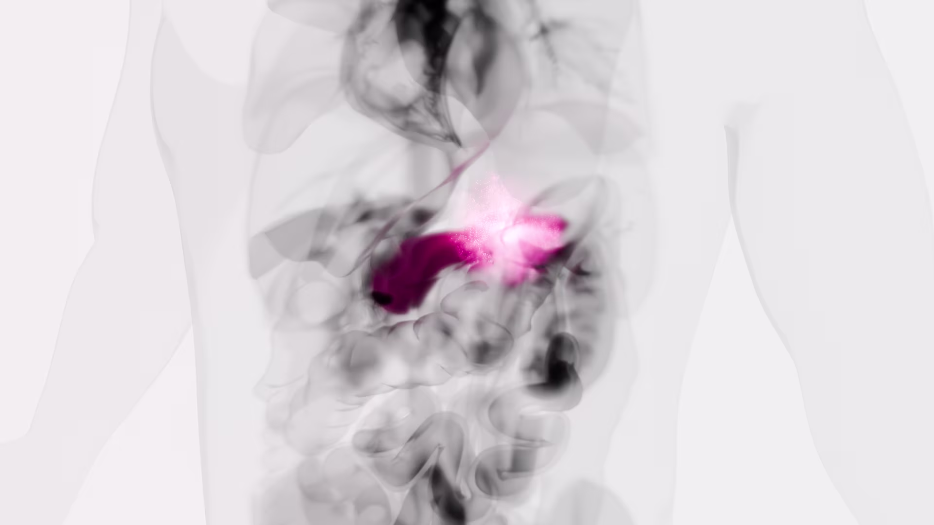
Pancreatic ductal adenocarcinoma (PDAC) is most often diagnosed in a metastatic stage. Circulating tumor cells (CTC) in the blood are hypothesized as the means of systemic dissemination. We aimed to isolate and characterize CTC to evaluate their significance as prognostic markers in PDAC. Blood obtained from healthy donors and patients with PDAC before therapy was filtered with ScreenCell® filtration devices for size-based CTC isolation. Captured cells were analyzed by immunofluorescence for an epithelial to mesenchymal transition (EMT) marker (zinc finger E-box binding homebox 1 (ZEB1)) and an epithelial antigen (cytokeratin (CK)). Molecular analysis of parallel specimens evaluated the KRAS mutation status of the CTC. The survival of each patient after study was recorded. As demonstrated by either cytology or finding of a KRAS mutation, CTC were detected in 18 of 21 patients (86 %) with proven PDAC: 8 out of 10 patients (80 %) with early stage (UICC IIA/IIB) and 10 out of 11 (91 %) with late stage (UICC III/IV) disease. CTC were not found in any of the 10 control patients (p < 0.001). The presence of CTC did not adversely affect median survival: 16 months in CTC-positive (n = 18) vs. 10 months in CTC-negative (n = 3) patients. Neither ZEB1 nor cytological characteristics correlated with overall survival, although ZEB1 was found almost exclusively in CTC of patients with established metastases. Patients with a CTC KRAS mutation (CTC-KRAS mut) had a substantially better survival, 19.4 vs. 7.4 months than patients with wild type KRAS (p = 0.015). With ScreenCell filtration, CTC are commonly found in PDAC (86 %). Molecular and genetic characterization, including mutations such as KRAS, may prove useful for prognosis.