
Cancer-associated Macrophage-like Cells in Patients with Non-metastatic Adenocarcinoma of the Esophagus – Cytomorphological Heterogeneity

Introduction: Esophageal adenocarcinoma (EAC) often recurs systemically despite therapy with a curative aim. New diagnostic and therapeutic approaches are urgently needed. A promising field is liquid biopsy, meaning the investigation of tumor-associated cells in the peripheral blood, for example cancer-associated macrophage-like cells (CAML). The aim of this multicentric study was to investigate the presence and cytomorphological appearance of CAML in patients with non-metastatic and operable esophageal cancer.
Methods: Blood samples from 252 patients with locally advanced EAC were obtained before starting curative treatment including surgery, and then processed using ScreenCell® filtration devices. Cytological analysis was performed via May-Grünwald-Giemsa staining. CAML were defined by their morphological characteristics. We also performed immunofluorescence staining with the mesenchymal marker vimentin on a subset of our study cohort.
Results: We detected cytomorphologically heterogeneous CAML in 31.8% (n=80) patients. Their presence and cell count did not correlate significantly with pretherapeutic cTNM. Even in patients with small tumors and no lymph-node infiltration, cell counts were high. CAML showed heterogenous staining patterns for vimentin.
Conclusion: This is one of the first studies demonstrating the presence and phenotype of CAML in a uniquely broad cohort of EAC patients. As they are believed to be representatives of the inflammatory tumor microenvironment shed into the bloodstream, their presence in non-metastatic EAC is a promising finding.
The Prognostic Value of the Circulating Tumor Cell-Based Four mRNA Scoring System: A New Non-Invasive Setting for the Management of Bladder Cancer

Simple Summary: Bladder cancer with similar diagnosis based on traditional classification exhibits different behaviors and therapeutic outcomes. Thus, circulating tumor cells (CTCs) represent a more accurate approach to investigate bladder cancer features. Our results demonstrate that risk score based on EGFR, TRPM4, TWIST1, and ZEB1 four-gene signature in CTCs is markedly and undoubtedly associated with recurrence, suggesting an innovative and non-invasive strategy to manage both non muscle invasive and muscle invasive bladder cancer progression without the
necessity of repetitive and onerous cystoscopies.
Abstract: Bladder cancer (BC) is one of the most expensive lifetime cancers to treat because of the high recurrence rate, repeated surgeries, and long-term cystoscopy monitoring and treatment. The lack of an accurate classification system predicting the risk of recurrence or progression leads to the search for new biomarkers and strategies. Our pilot study aimed to identify a prognostic gene signature in circulating tumor cells (CTCs) isolated by ScreenCell devices from muscle invasive and non-muscle invasive BC patients. Through the PubMed database and Cancer Genome Atlas dataset, a panel
of 15 genes modulated in BC with respect to normal tissues was selected. Their expression was evaluated in CTCs and thanks to the univariate and multivariate Cox regression analysis, EGFR, TRPM4, TWIST1, and ZEB1 were recognized as prognostic biomarkers. Thereafter, by using the risk score model, we demonstrated that this 4-gene signature significantly grouped patients into high- and low-risk in terms of recurrence free survival (HR = 2.704, 95% CI = 1.010–7.313, Log-rank p < 0.050). Overall, we identified a new prognostic signature that directly impacted the prediction of recurrence, improving the choice of the best treatment for BC patients.
Identification of Atypical Circulating Tumor Cells with Prognostic Value in Metastatic Breast Cancer Patients

Circulating tumor cells have a strong potential as a quasi-non-invasive tool for setting up a precision medicine strategy for cancer patients. Using a second-generation « »filtration-based » » technology to isolate CTCs, the Screencell™ technology (Sarcelles, France), we performed a large and simultaneous analysis of all atypical circulating tumor cells (aCTCs) isolated from the blood of metastatic breast cancer (mBC) patients. We correlated their presence with clinicopathological and survival data. We included 91 mBC patients from the PERMED-01 study. The median number of aCTCs was 8.3 per mL of blood. Three subsets of aCTCs, absent from controls, were observed in patients: single (s-aCTCs), circulating tumor micro-emboli (CTM), and giant-aCTCs (g-aCTCs). The presence of g-aCTCs was associated with shorter progression free survival and overall survival. This study highlights the heterogeneity of aCTCs in mBC patients both at the cytomorphological and molecular levels. In addition, it suggests the usefulness of the g-aCTC subset as a prognostic factor and a potential stratification tool to treat late-stage mBC patients and improve their chances of benefiting from early clinical trials.
Sequential Isolation and Characterization of Single CTCs and Large CTC Clusters in Metastatic Colorectal Cancer Patients

Circulating tumor cells (CTCs) detach from a primary tumor or its metastases and circulate in the bloodstream. The vast majority of CTCs are deemed to die into the bloodstream, with only few cells representing viable metastatic precursors. Particularly, single epithelial CTCs do not survive long in the circulation due to the loss of adhesion-dependent survival signals. In metastatic colorectal cancer, the generation of large CTC clusters is a very frequent occurrence, able to increase the aptitude of CTCs to survive in the bloodstream. Although a deepened analysis of large-sized CTC clusters might certainly offer new insights into the complexity of the metastatic cascade, most CTC isolation techniques are unfortunately not compatible with large-sized CTC clusters isolation. The inappropriateness of standard CTC isolation devices for large clusters isolation and the scarce availability of detection methods able to specifically isolate and characterize both single CTCs and CTC clusters finally prevented in-depth studies on the prognostic and predictive value of clusters in clinical practice, unlike that which has been described for single CTCs. In the present study, we validated a new sequential filtration method for the simultaneous isolation of large CTC clusters and single CTCs in patients with metastatic colorectal cancer at failure of first-line treatments. The new method might allow differential downstream analyses for single and clustered CTCs starting from a single blood draw, opening new scenarios for an ever more precise characterization of colorectal cancer metastatic cascade.
Circulating Tumor Cell Clusters Are Frequently Detected in Women with Early-Stage Breast Cancer
The clinical relevance of circulating tumor cell clusters (CTC-clusters) in breast cancer (BC) has been mostly studied using the CellSearch®, a marker-dependent method detecting only epithelial-enriched clusters. However, due to epithelial-to-mesenchymal transition, resorting to marker-independent approaches can improve CTC-cluster detection. Blood samples collected from healthy donors and spiked-in with tumor mammospheres, or from BC patients, were processed for CTC-cluster detection with 3 technologies: CellSearch®, CellSieve™ filters, and ScreenCell® filters. In spiked-in samples, the 3 technologies showed similar recovery capability, whereas, in 19 clinical samples processed in parallel with CellSearch® and CellSieve™ filters, filtration allowed us to detect more CTC-clusters than CellSearch® (median number = 7 versus 1, p = 0.0038). Next, samples from 37 early BC (EBC) and 23 metastatic BC (MBC) patients were processed using ScreenCell® filters for attaining both unbiased enrichment and marker-independent identification (based on cytomorphological criteria). At baseline, CTC-clusters were detected in 70% of EBC cases and in 20% of MBC patients (median number = 2, range 0-20, versus 0, range 0-15, p = 0.0015). Marker-independent approaches for CTC-cluster assessment improve detection and show that CTC-clusters are more frequent in EBC than in MBC patients, a novel finding suggesting that dissemination of CTC-clusters is an early event in BC natural history.
Isolation and Enumeration of CTC in Colorectal Cancer Patients: Introduction of a Novel Cell Imaging Approach and Comparison to Cellular and Molecular Detection Techniques
Circulating tumour cells (CTC) were proven to be prognostically relevant in cancer treatment, e.g., in colorectal cancer (CRC). This study validates a molecular detection technique through using a novel cell imaging approach for CTC detection and enumeration, in comparison to a size-based cellular and correlated the data to clinico-pathological characteristics. Overall, 57 CRC patients were recruited for this prospective study. Blood samples were analysed for CTCs by three methods: (1) Epithelial marker immunofluorescence staining combined with automated microscopy using the NYONE® cell imager; (2) isolation by size using membrane filtration with the ScreenCell® Cyto IS device and immunofluorescence staining; (3) detection by semi-quantitative Cytokeratin-20 RT-qPCR. Enumeration data were compared and correlated with clinic-pathological parameters. CTC were detected by either approach; however, with varying positivity rates: NYONE® 36.4%, ScreenCell® 100%, and PCR 80.5%. All methods revealed a positive correlation of CTC presence and higher tumour burden, which was most striking using the ScreenCell® device. Generally, no intercorrelation of CTC presence emerged amongst the applied techniques. Overall, enumeration of CTC after isolation by size demonstrated to be the most reliable strategy for the detection of CTC in CRC patients. Ongoing studies will have to unravel the prognostic value of this finding, and validate this approach in a larger cohort.
Comparative performance of different methods for circulating tumor cell enrichment in metastatic breast cancer patients
The isolation and analysis of circulating tumor cells (CTC) has the potential to provide minimally invasive diagnostic, prognostic and predictive information. Widespread clinical implementation of CTC analysis has been hampered by a lack of comparative investigation between different analytic methodologies in clinically relevant settings. The objective of this study was to evaluate four different CTC isolation techniques-those that rely on surface antigen expression (EpCAM or CD45 using DynaBeads® or EasySep™ systems) or the biophysical properties (RosetteSep™ or ScreenCell®) of CTCs. These were evaluated using cultured cells in order to calculate isolation efficiency at various levels including; inter-assay and inter-operator variability, protocol complexity and turn-around time. All four techniques were adequate at levels above 100 cells/mL which is commonly used for the evaluation of new isolation techniques. Only the RosetteSep™ and ScreenCell® techniques were found to provide adequate sensitivity at a level of 10 cells/mL. These techniques were then applied to the isolation and analysis of circulating tumor cells blood drawn from metastatic breast cancer patients where CTCs were detected in 54% (15/28) of MBC patients using the RosetteSep™ and 75% (6/8) with ScreenCell®. Overall, the ScreenCell® method had better sensitivity.
Detection of circulating tumor cells in patients with laryngeal cancer using ScreenCell: Comparative pre- and post-operative analysis and association with prognosis
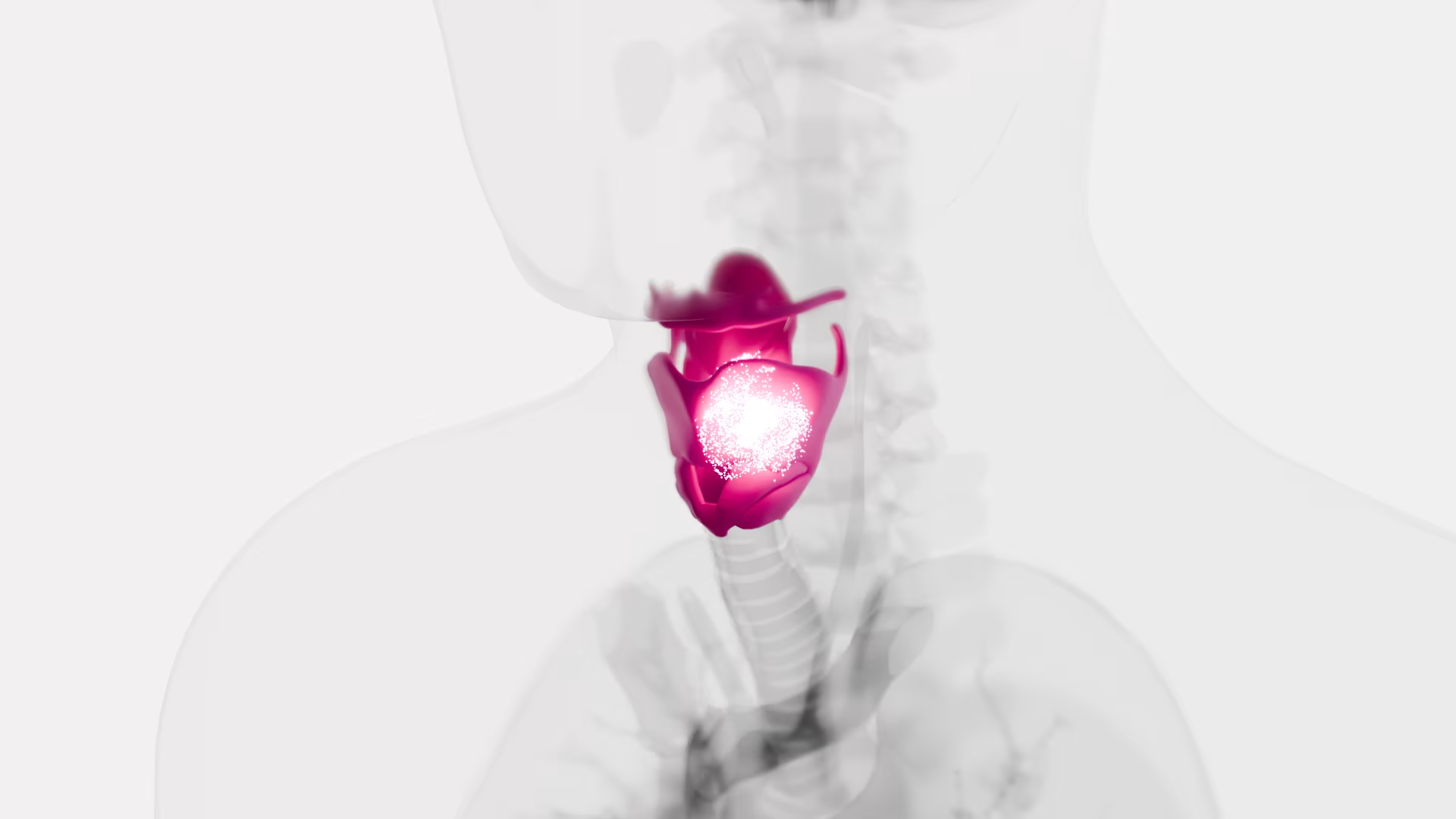
The presence of circulating tumor cells (CTCs) in the blood of patients with metastatic breast, colorectal and prostate cancer have been widely investigated; however, few studies have examined CTCs in patients with laryngeal cancer. The present pilot study aimed to detect pre- and postoperative CTCs in the blood of patients with laryngeal cancer and evaluate the association with prognosis. Eight patients with laryngeal squamous cell carcinoma (LSCC) at stage III were included in the present study and underwent total or subtotal laryngectomy and radical bilateral neck lymph node dissection. Blood samples were collected from all patients before and after surgery at different time-points. The following processing steps were followed; preoperative blood sampling, surgery, postoperative blood sampling at 3, 6 and 12 month follow-ups, and prognostic association analysis. CTCs were retained on ScreenCell filters for cytological characterization. The presence of CTCs was associated with a less favorable prognosis, whereas a decrease of CTCs in the postoperative sampling was observed in patients who exhibited an improved therapeutic response. The results of the present pilot study revealed a possible association between the presence of CTCs and a less favorable prognosis in patients with LSCC; therefore, these preliminary findings may encourage further research into the incorporation of a liquid biopsy in the management of LSCC, as this may help identify patients with occult metastatic disease earlier and in a non-invasive manner. In addition, this approach may represent novel independent prognostic factor for use in the clinical evaluation of patients with LSCC.
Circulating Tumor Cells in Right- and Left-Sided Colorectal Cancer
Molecular alterations are not randomly distributed in colorectal cancer (CRC), but rather clustered on the basis of primary tumor location underlying the importance of colorectal cancer sidedness. We aimed to investigate whether circulating tumor cells (CTC) characterization might help clarify how different the patterns of dissemination might be relative to the behavior of left- (LCC) compared to right-sided (RCC) cancers. We retrospectively analyzed patients with metastatic CRC who had undergone standard baseline CTC evaluation before starting any first-line systemic treatment. Enumeration of CTC in left- and right-sided tumors were compared. The highest prognostic impact was exerted by CTC in left-sided primary cancer patients, even though the lowest median number of cells was detected in this subgroup of patients. CTC exhibit phenotypic heterogeneity, with a predominant mesenchymal phenotype found in CTC from distal compared to proximal primary tumors. Most CTC in RCC patients exhibited an apoptotic pattern. CTC in left-sided colon cancer patients exhibit a predominant mesenchymal phenotype. This might imply a substantial difference in the biology of proximal and distal cancers, associated with different patterns of tumor cells dissemination. The poor prognosis of right-sided CRC is not determined by the hematogenous dissemination of tumor cells, which appears to be predominantly a passive shedding of non-viable cells. Conversely, the subgroup of poor-prognosis left-sided CRC is reliably identified by the presence of mesenchymal CTC.
Prognostic Relevance of Circulating Tumor Cells and Circulating Cell-Free DNA Association in Metastatic Non-Small Cell Lung Cancer Treated with Nivolumab

The treatment of advanced non-small cell lung cancer (NSCLC) has been revolutionized by immune checkpoint inhibitors (ICIs). The identification of prognostic and predictive factors in ICIs-treated patients is presently challenging. Circulating tumor cells (CTCs) and cell-free DNA (cfDNA) were evaluated in 89 previously treated NSCLC patients receiving nivolumab. Blood samples were collected before therapy and at the first and second radiological response assessments. CTCs were isolated by a filtration-based method. cfDNA was extracted from plasma and estimated by quantitative PCR. Patients with baseline CTC number and cfDNA below their median values (2 and 836.5 ng from 3 mL of blood and plasma, respectively) survived significantly longer than those with higher values (p = 0.05 and p = 0.04, respectively). The two biomarkers were then used separately and jointly as time-dependent covariates in a regression model confirming their prognostic role. Additionally, a four-fold risk of death for the subgroup presenting both circulating biomarkers above the median values was observed (p < 0.001). No significant differences were found between circulating biomarkers and best response. However, progressing patients with concomitant lower CTCs and cfDNA performed clinically well (p = 0.007), suggesting that jointed CTCs and cfDNA might help discriminate a low-risk population which might benefit from continuing ICIs beyond progression.