
Weighted correlation network analysis revealed novel long non-coding RNAs for colorectal cancer
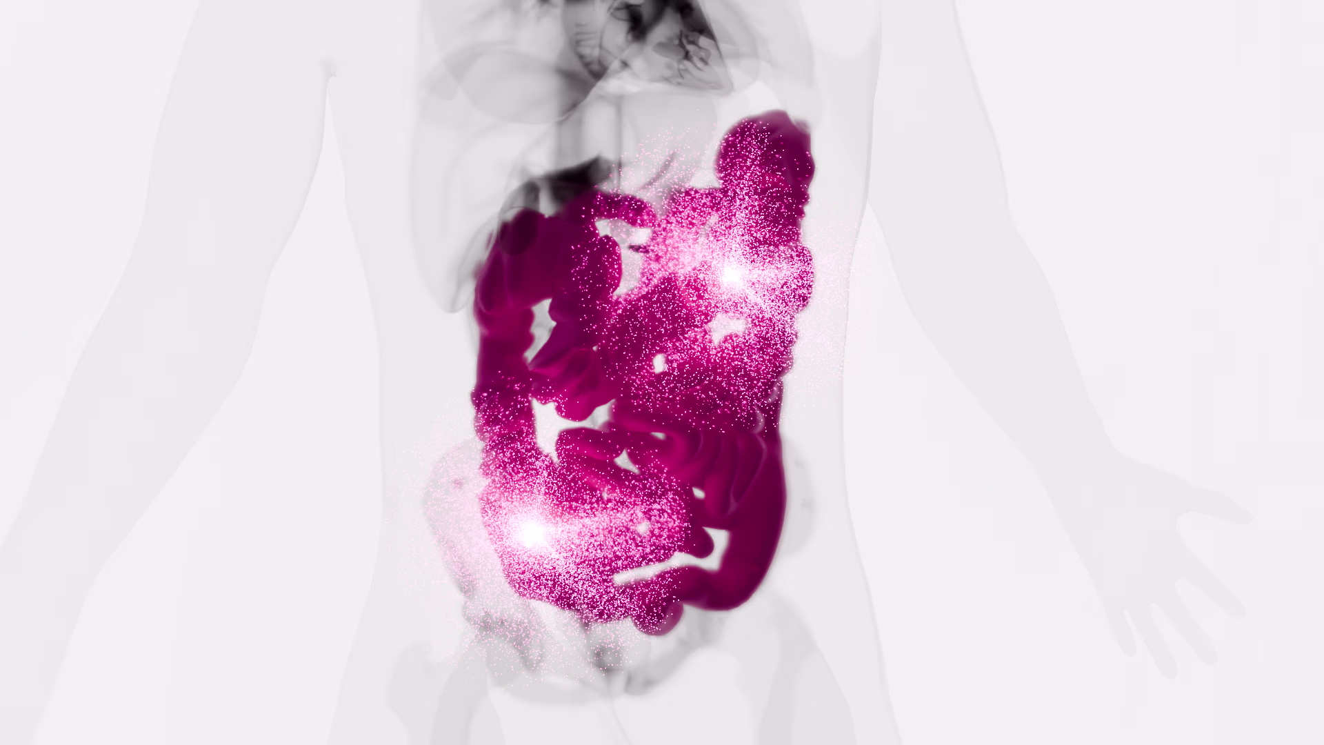
Colorectal cancer (CRC) is one of the most prevalent cancers worldwide, which after breast, lung and, prostate cancers, is the fourth prevalent cancer in the United States. Long non-coding RNAs (lncRNAs) have an essential role in the pathogenesis of CRC. Therefore, bioinformatics studies on lncRNAs and their target genes have potential importance as novel biomarkers. In the current study, publicly available microarray gene expression data of colorectal cancer (GSE106582) was analyzed with the Limma, Geoquery, Biobase package. Afterward, identified differentially expressed lncRNAs and their target genes were inserted into Weighted correlation network analysis (WGCNA) to obtain modules and hub genes. A total of nine differentially expressed lncRNAs (LINC01018, ITCH-IT, ITPK1-AS1, FOXP1-IT1, FAM238B, PAXIP1-AS1, ATP2B1-AS1, MIR29B2CHG, and SNHG32) were identified using microarray data analysis. The WGCNA has identified several hub genes for black (LMOD3, CDKN2AIPNL, EXO5, ZNF69, BMS1P5, METTL21A, IL17RD, MIGA1, CEP19, FKBP14), blue (CLCA1, GUCA2A, UGT2B17, DSC2, CA1, AQP8, ITLN1, BEST4, KLF4, IQCF6) and turquoise (PAFAH1B1, LMNB1, CACYBP, GLO1, PUM3, POC1A, ASF1B, SDCCAG3, ASNS, PDCD2L) modules. The findings of the current study will help to improve our understanding of CRC. Moreover, the hub genes that we have identified could be considered as possible prognostic/diagnostic biomarkers. This study led to the determination of nine lncRNAs with no previous association with CRC development.
Sequential Isolation and Characterization of Single CTCs and Large CTC Clusters in Metastatic Colorectal Cancer Patients
Circulating tumor cells (CTCs) detach from a primary tumor or its metastases and circulate in the bloodstream. The vast majority of CTCs are deemed to die into the bloodstream, with only few cells representing viable metastatic precursors. Particularly, single epithelial CTCs do not survive long in the circulation due to the loss of adhesion-dependent survival signals. In metastatic colorectal cancer, the generation of large CTC clusters is a very frequent occurrence, able to increase the aptitude of CTCs to survive in the bloodstream. Although a deepened analysis of large-sized CTC clusters might certainly offer new insights into the complexity of the metastatic cascade, most CTC isolation techniques are unfortunately not compatible with large-sized CTC clusters isolation. The inappropriateness of standard CTC isolation devices for large clusters isolation and the scarce availability of detection methods able to specifically isolate and characterize both single CTCs and CTC clusters finally prevented in-depth studies on the prognostic and predictive value of clusters in clinical practice, unlike that which has been described for single CTCs. In the present study, we validated a new sequential filtration method for the simultaneous isolation of large CTC clusters and single CTCs in patients with metastatic colorectal cancer at failure of first-line treatments. The new method might allow differential downstream analyses for single and clustered CTCs starting from a single blood draw, opening new scenarios for an ever more precise characterization of colorectal cancer metastatic cascade.
Isolation and Enumeration of CTC in Colorectal Cancer Patients: Introduction of a Novel Cell Imaging Approach and Comparison to Cellular and Molecular Detection Techniques
Circulating tumour cells (CTC) were proven to be prognostically relevant in cancer treatment, e.g., in colorectal cancer (CRC). This study validates a molecular detection technique through using a novel cell imaging approach for CTC detection and enumeration, in comparison to a size-based cellular and correlated the data to clinico-pathological characteristics. Overall, 57 CRC patients were recruited for this prospective study. Blood samples were analysed for CTCs by three methods: (1) Epithelial marker immunofluorescence staining combined with automated microscopy using the NYONE® cell imager; (2) isolation by size using membrane filtration with the ScreenCell® Cyto IS device and immunofluorescence staining; (3) detection by semi-quantitative Cytokeratin-20 RT-qPCR. Enumeration data were compared and correlated with clinic-pathological parameters. CTC were detected by either approach; however, with varying positivity rates: NYONE® 36.4%, ScreenCell® 100%, and PCR 80.5%. All methods revealed a positive correlation of CTC presence and higher tumour burden, which was most striking using the ScreenCell® device. Generally, no intercorrelation of CTC presence emerged amongst the applied techniques. Overall, enumeration of CTC after isolation by size demonstrated to be the most reliable strategy for the detection of CTC in CRC patients. Ongoing studies will have to unravel the prognostic value of this finding, and validate this approach in a larger cohort.
Circulating Tumor Cells in Right- and Left-Sided Colorectal Cancer
Molecular alterations are not randomly distributed in colorectal cancer (CRC), but rather clustered on the basis of primary tumor location underlying the importance of colorectal cancer sidedness. We aimed to investigate whether circulating tumor cells (CTC) characterization might help clarify how different the patterns of dissemination might be relative to the behavior of left- (LCC) compared to right-sided (RCC) cancers. We retrospectively analyzed patients with metastatic CRC who had undergone standard baseline CTC evaluation before starting any first-line systemic treatment. Enumeration of CTC in left- and right-sided tumors were compared. The highest prognostic impact was exerted by CTC in left-sided primary cancer patients, even though the lowest median number of cells was detected in this subgroup of patients. CTC exhibit phenotypic heterogeneity, with a predominant mesenchymal phenotype found in CTC from distal compared to proximal primary tumors. Most CTC in RCC patients exhibited an apoptotic pattern. CTC in left-sided colon cancer patients exhibit a predominant mesenchymal phenotype. This might imply a substantial difference in the biology of proximal and distal cancers, associated with different patterns of tumor cells dissemination. The poor prognosis of right-sided CRC is not determined by the hematogenous dissemination of tumor cells, which appears to be predominantly a passive shedding of non-viable cells. Conversely, the subgroup of poor-prognosis left-sided CRC is reliably identified by the presence of mesenchymal CTC.
EpCAM-expressing circulating tumor cells in colorectal cancer
Background: Several studies have raised the issue of the inadequacy of CellSearch® to detect the entire pool of circulating tumor cells (CTCs) from blood of cancer patients, suggesting that cells expressing low levels of epithelial cell adhesion molecule (EpCAM) are not recognized by the capture reagent. In this exploratory study, we aimed to evaluate the status of EpCAM in CTCs isolated from a group of metastatic colorectal cancer patients, in 40% of whom, CTC had been found to be undetected by the CellSearch® system.
Methods: CTCs were analyzed using both a microfiltration method (ScreenCell) and CellSearch® in parallel. Furthermore, since EpCAM exists in 2 different variants, we investigated the presence of both its intracellular domain (EpICD) and extracellular domain (EpEX) through immunofluorescence staining of CTCs on filters.
Results: Results from immunofluorescence experiments demonstrated that, overall, EpICD and/or EpEX was expressed in 176 CTCs detected by ScreenCell, while the CellSearch® system was able to capture only 10 CTCs.
Conclusions: This is the first demonstration that the low sensitivity of CellSearch® to detect CTCs in colorectal cancer patients is not due to the lack of EpCAM. »
Détection des mutations RAS dans les cellules tumorales circulantes : applications au cancer colorectal et perspectives
Les mutations somatiques présentes dans les gènes RAS (KRAS et NRAS) sont largement associées à l’absence de réponse aux traitements par immunothérapie ciblant le récepteur du facteur de croissance épidermique dans le cancer colorectal métastatique. La recherche de ces mutations est devenue obligatoire pour pouvoir prescrire ces traitements et cette détection est réalisée à partir de biopsies tissulaires. Dans environ 25 % des cas, cette analyse n’est pas possible pour des raisons à la fois analytique et médicale et par conséquent le développement de stratégies alternatives est donc nécessaire. Parmi les solutions envisagées, la recherche de mutations directement dans le sang des patients est une approche prometteuse. Parmi les sources d’ADN tumoral présent dans la circulation sanguine, les cellules tumorales circulantes (CTC) représentent une approche particulièrement pertinente. Ces cellules, dont certaines sont à l’origine des métastases, sont parvenues à se détacher de la tumeur primitive, puis à migrer et enfin à entrer dans le système vasculaire. En ce sens, elles sont particulièrement résistantes aux contraintes physico-chimiques et immunologiques mises en œuvre par l’organisme pour empêcher leur dissémination et représentent par conséquent une source d’informations particulièrement précieuse sur la génétique des cellules tumorales les plus agressives. Le corollaire est que ces cellules sont très rares et nécessitent des technologies particulièrement performantes pour les détecter et les caractériser. Dans cette présentation, nous nous focaliserons principalement sur les méthodes moléculaires permettant de détecter les mutations des gènes RAS sur les CTC en analysant les performances d’une solution basée sur une méthode d’enrichissement par filtration suivi d’une détection par PCR digitale. Enfin, nous nous interrogerons sur leur signification biologique avant d’évoquer leurs perspectives dans le cancer colorectal ainsi que dans d’autres types de cancers.
Droplet digital PCR of circulating tumor cells from colorectal cancer patients can predict KRAS mutations before surgery
In colorectal cancer (CRC), KRAS mutations are a strong negative predictor for treatment with the EGFR-targeted antibodies cetuximab and panitumumab. Since it can be difficult to obtain appropriate tumor tissues for KRAS genotyping, alternative methods are required. Circulating tumor cells (CTCs) are believed to be representative of the tumor in real time. In this study we explored the capacity of a size-based device for capturing CTCs coupled with a multiplex KRAS screening assay using droplet digital PCR (ddPCR). We showed that it is possible to detect a mutant ratio of 0.05% and less than one KRAS mutant cell per mL total blood with ddPCR compared to about 0.5% and 50–75 cells for TaqMeltPCR and HRM. Next, CTCs were isolated from the blood of 35 patients with CRC at various stage of the disease. KRAS genotyping was successful for 86% (30/35) of samples with a KRAS codon 12/13 mutant ratio of 57% (17/30). In contrast, only one patient was identified as KRAS mutant when size-based isolation was combined with HRM or TaqMeltPCR. KRAS status was then determined for the 26 available formalin-fixed paraffin-embedded tumors using standard procedures. The concordance between the CTCs and the corresponding tumor tissues was 77% with a sensitivity of 83%. Taken together, the data presented here suggest that is feasible to detect KRAS mutations in CTCs from blood samples of CRC patients which are predictive for those found in the tumor. The minimal invasive nature of this procedure in combination with the high sensitivity of ddPCR might provide in the future an opportunity to monitor patients throughout the course of disease on multiple levels including early detection, prognosis, treatment and relapse as well as to obtain mechanistic insight with respect to tumor invasion and metastasis.
Colorectal carcinomas in 2013: the search for powerful prognostic markers is still on the go!
Colorectal cancer (CRC) is the third cause of cancer worldwide after prostate cancer and breast cancer. Patients have a survival rate of 5 years, which varies between 10 and 95% depending on CRC stage. Today, the management of patients with CRC is based on parameters such as TNM and classic histologic parameters, but new molecular and cell markers have been created to improve treatment and survival. Determining the expression of a characteristic set of genes either from formalin-fixed paraffin-embedded tissue (Onco type DX test™) or from fresh tissues (AGENDIA© ColoPrint®) has led to encouraging results, but there is a need for clinical validation on a large number of patients. Also, next-generation sequencing (NGS) technologies may be the next step in the molecular approach of CRC tumor samples, allowing tumor characterization by gene signature arrays. In addition to molecular markers, evaluation of the presence of cellular markers such as circulating tumor cells (CTC) in the blood of patients with CRC can optimize prognostic evaluation and response to treatment. CTC isolation methods used today have different sensitivities and specificities, due not only to the very small number of these cells but also to the epithelial-mesenchymal transitional process (EMT). This paper presents the preliminary results of our study conducted on CTC isolation in patients with CRC by filtration method (Screencells Cyto®). This fast and efficient method identifies CTCs and also isolates cells in EMT, which explains its high efficiency compared to technologies based on immunomagnetic and microfluidic separation reliant on EpCAM presence on the cell surface.